

Case Reports
doi: 10.1002/hep.27872.
Epub 2015 Jun 19.
Hepatocellular carcinoma associated with tight-junction protein 2 deficiency
Affiliations
- PMID: 25921221
- PMCID: PMC4626433
- DOI: 10.1002/hep.27872
Item in Clipboard
Case Reports
Hepatocellular carcinoma associated with tight-junction protein 2 deficiency
Hepatology.
2015 Dec.
No abstract available
Figures

Patient 1. Non-neoplastic liver shows micronodular cirrhosis (a) and prominent rosetting with bile plugs in canalicular lumina (b). c-d. Low and high-magnification views of HCC. Tumor cells show variable cytoplasmic clearing and round to oval, sometimes pleomorphic nuclei with variably-sized nucleoli. Some cells contain distinct round to oval eosinophilic cytoplasmic proteinaceous inclusions (arrow in d). Macrosteatosis and mitosis (arrow in e) are also present. Bile salt export pump (f) and multiple drug resistance protein 3 (g) are well-expressed in nonneoplastic liver. h. Complete absence of tight junction protein 2 (TJP2) staining in bile ducts. i. Markedly decreased claudin-1 staining in both cholangiocytes and canalicular margins. “Control liver” shows normal TJP2 expression in bile duct (inset in h) and strong claudin-1 expression in both bile ducts and canaliculi (inset in i). (a. Trichrome stain; b-f. Hematoxylin-eosin stain; Original magnification: 40 × for a, 100 × for c, 200 × for b, d and e, 400 × for f-i)
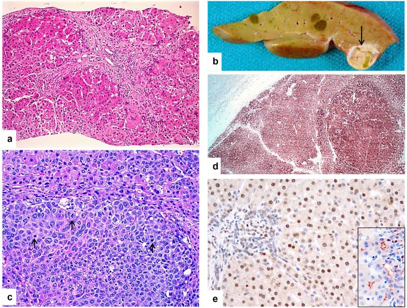
Patient 2. a. Liver biopsy at 6 months displays severe cholestasis, giant cell transformation, and micronodular cirrhosis. b. Macroscopic photograph of the explant reveals multiple cholestatic nodules, and one much larger and softer tan nodule with focal bile pigmentation (arrow) within which hepatocellular carcinoma (HCC) was found histologically. c. Within the large, soft nodule, a sharply delineated smaller aggregate of highly pleomorphic and mitotically active cells (arrows) is typical of HCC. d. Glypican-3 expression (brown staining) is seen in HCC and regenerative nodule but not in non-neoplastic hepatocytes (left upper). e. Tight junction protein 2 (TJP2) is not expressed at bile canaliculi of the patient's liver-biopsy specimen, whereas it is observed anomalously in hepatocyte nuclei. “Control liver” shows normal canalicular expression of TJP2 (inset). (a & c: Hematoxylin-eosin stain; Original magnification: 125 × for d, 200 × for a, c and e)
References
-
- Knisely AS, Strautnieks SS, Meier Y, Stieger B, Byrne JA, Portmann BC, et al. Hepatocellular carcinoma in ten children under five years of age with bile salt export pump deficiency. Hepatology. 2006;44:478–486. - PubMed
-
- Gonzalez-Mariscal L, Lechuga S, Garay E. Role of tight junctions in cell proliferation and cancer. Progress in Histochemistry and Cytochemistry. 2007;42:1–57. - PubMed
-
- Vilarinho S, Erson-Omay EZ, Harmanci AS, Morotti R, Carrion-Grant G, Baranoski J, et al. Paediatric hepatocellular carcinoma due to somatic CTNNB1 and NFE2L2 mutations in the setting of inherited bi-allelic ABCB11 mutations. Journal of Hepatology. 2014;61:1178–1183. - PubMed
-
- Iannelli F, Collino A, Sinha S, Radaelli E, Nicoli P, D'Antiga L, et al. Massive gene amplification drives paediatric hepatocellular carcinoma caused by bile salt export pump deficiency. Nature Communications. 2014;5:3850. - PubMed
Publication types
MeSH terms
Substances
Grants and funding
LinkOut - more resources
Full Text Sources
Other Literature Sources
Medical