Clinical Trial
. 2016 Jan;9(1):56-67.
doi: 10.1038/mi.2015.35. Epub 2015 Apr 29.

Modulation of nasopharyngeal innate defenses by viral coinfection predisposes individuals to experimental pneumococcal carriage

Affiliations
Clinical Trial

Modulation of nasopharyngeal innate defenses by viral coinfection predisposes individuals to experimental pneumococcal carriage

S Glennie et al. Mucosal Immunol. 2016 Jan.

Abstract

Increased nasopharyngeal colonization density has been associated with pneumonia. We used experimental human pneumococcal carriage to investigate whether upper respiratory tract viral infection predisposes individuals to carriage. A total of 101 healthy subjects were screened for respiratory virus before pneumococcal intranasal challenge. Virus was associated with increased odds of colonization (75% virus positive became colonized vs. 46% virus-negative subjects; P=0.02). Nasal Factor H (FH) levels were increased in virus-positive subjects and were associated with increased colonization density. Using an in vitro epithelial model we explored the impact of increased mucosal FH in the context of coinfection. Epithelial inflammation and FH binding resulted in increased pneumococcal adherence to the epithelium. Binding was partially blocked by antibodies targeting the FH-binding protein Pneumococcal surface protein C (PspC). PspC epitope mapping revealed individuals lacked antibodies against the FH binding region. We propose that FH binding to PspC in vivo masks this binding site, enabling FH to facilitate pneumococcal/epithelial attachment during viral infection despite the presence of anti-PspC antibodies. We propose that a PspC-based vaccine lacking binding to FH could reduce pneumococcal colonization, and may have enhanced protection in those with underlying viral infection.


Conflict of interest statement

The authors declared no conflict of interest.

Figures

Figure 1
Figure 1
Asymptomatic upper respiratory tract (URT) viral infections are associated with susceptibility to pneumococcal colonization but not increased density. (a) A total of 101 healthy subjects were screened for URT viruses by multiplex PCR 5 days before intranasal inoculation of 6B pneumococcus. The percentage of carriage-positive (carriers) and carriage-negative (noncarriers) subjects are shown for virus negative or virus positive. Among virus-positive subjects, 75% became colonized as compared with 46% virus-negative subjects (P=0.02, using Fisher’s exact test). (b) Colonization density recovered from the nasopharynx was measured in nasal wash samples collected 2, 7, and 14 days following pneumococcal inoculation and is expressed as CFU ml−1 recovered from nasal wash (NW) of virus-negative (no virus) and virus-positive (virus) groups. There was no difference in colonization density between virus-positive and virus-negative subjects. CFU, colony-forming unit; neg, negative; pos, positive. PowerPoint slide
Figure 2
Figure 2
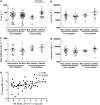
Levels of mucosal Factor H (FH) are increased in individuals coinfected with virus and pneumococci and correlate with colonization density. Mucosal levels of (a) Factor H, (b) secretory leukocyte proteinase inhibitor (SLPI), (c) β-defensin 2, and (d) lactoferrin were measured in nasal wash samples obtained 2 days after pneumococcal challenge with 6B. Levels are expressed in ng ml−1 and results are stratified by pneumococcal carriage and absence or presence of virus. Statistical differences were analyzed using unpaired Student’s t-test (*P≤0.05). (e) We observed a positive correlation between levels of FH and pneumococcal colonization density (CFU ml−1) recovered from nasal wash (NW) 2 days after inoculation using Pearson’s correlation test. CFU, colony-forming unit. P-value and the correlation coefficient (r) are presented. PowerPoint slide
Figure 3
Figure 3
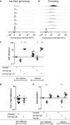
Pneumococcal epithelial adherence and internalization are increased in the presence of human nasal wash (NW) or Factor H (FH). Adherence of (a) D39 and (b) D39ΔPspC to human pharyngeal epithelial cells (Detroit 562) was evaluated in noninflamed and inflamed epithelium. PspC, Pneumococcal surface protein C. (a) Epithelial adherence and (c) internalization of pneumococci was increased following treatment of bacteria with human purified FH (open dots), NW depleted of IgA (gray dots), and NW depleted of IgG (black dots) when compared with untreated bacteria (black squares). (b) Epithelial adherence was not increased when D39ΔPspC was pretreated with FH (open dots). All conditions were performed in duplicate and experiments were repeated at least twice. For each experiment, the average recovered colony-forming units (CFUs) from the control condition (adherence of D39 untreated bacteria to noninflamed cells) was used to calculate fold change of all remaining conditions. The symbol + represents statistical significance compared with control condition using unpaired t-test (+P≤0.05 and ++P≤0.005). The symbol * represents statistical significance compared with untreated bacteria of same epithelial condition using unpaired Student’s t-test (*P≤0.05 and **P≤0.005). PowerPoint slide
Figure 4
Figure 4
Purified human anti-PspC immunoglobulin G (IgG) binds to pneumococcus and partially inhibits Factor H (FH) binding and adherence to human pharyngeal epithelial cells. (a) We measured anti-PspC IgG binding to D39 and (b) inhibition of FH binding by purified anti-PspC IgG. PspC, Pneumococcal surface protein C. Each row represents one subject. Negative controls (without anti-PspC or FH) are represented by unfilled histograms (a, b). For the inhibition of FH assay the positive control (without anti-PspC and with FH) is represented by black filled histogram. (c) Epithelial adherence of D39 was evaluated following treatment of bacteria with purified anti-PspC IgG containing low levels of FH (3.5 μg ml−1 residual from purification of serum samples, gray dots) or with anti-PspC IgG before addition of high levels of FH (10 μg ml−1, open dots) and compared with untreated bacteria. (d) Adherence of D39ΔPspC untreated (black squares) or treated with purified anti-PspC IgG (gray dots) was evaluated. (e) D39 epithelial internalization was measured in untreated bacteria (black squares) and bacteria treated with the purified anti-PspC IgG (gray dots) or the total IgG purified from the same serum samples as control for antibody specificity (open squares). The symbol + represents statistical significance compared with control condition using unpaired t-test (+P≤0.05 and ++P≤0.01). The symbol ** represents statistical significance compared with untreated bacteria on inflamed epithelial condition (black squares) using unpaired t-test (*P≤0.05 and **P≤0.005). PowerPoint slide
Figure 5
Figure 5
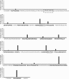
Pneumococcal surface protein C (PspC) epitope mapping using sera of healthy adults exposed to pneumococcus reveals the lack of antibodies to the Factor H (FH) binding site. The amino acid sequence of PspC group 3 variant was spotted on peptide arrays and is represented on the x axis. Peptide arrays covering the N-terminal region until the proline-rich region were probed with 29 samples from 18 subjects. The number of samples that recognized each peptide is represented on the y axis. Sequences of the peptides recognized by human sera are in black and the ones not recognized are in gray. Binding sites for FH and secretory immunoglobulin A (sIgA) are represented in dashed black and continuous gray boxes, respectively. PowerPoint slide
Figure 6
Figure 6
Schematic model of the proposed relationship between Pneumococcal surface protein C (PspC), Factor H (FH), anti-PspC immunoglobulin G (IgG), and pneumococcal adherence. (Left column) Untreated condition—no FH or other factors such as vitronectin are present. Adherence is moderate. (Middle column) In the presence of anti-PspC and low levels of FH there is decreased adherence because anti-PspC antibodies block pneumococcal adherence to the epithelium mediated by other factors, such as vitronectin. Human anti-PspC antibodies do not recognize the FH binding site of PspC and therefore the FH–PspC interaction with continue to facilitate low levels of pneumococcal adherence. (Right column) In the presence of anti-PspC and high levels of FH there is increased adherence because of increased interactions of PspC–FH. PowerPoint slide

References

    1. Morens DM, Taubenberger JK, Fauci AS. Predominant role of bacterial pneumonia as a cause of death in pandemic influenza: implications for pandemic influenza preparedness. J. Infect. Dis. 2008;198:962–970. doi: 10.1086/591708. - DOI - PMC - PubMed
    1. McCullers JA. Influenza enhances susceptibility to natural acquisition of and disease due to Streptococcus pneumoniae in ferrets. J. Infect. Dis. 2010;202:1287–1295. doi: 10.1086/656333. - DOI - PMC - PubMed
    1. Vu HT. Association between nasopharyngeal load of Streptococcus pneumoniae, viral coinfection, and radiologically confirmed pneumonia in Vietnamese children. Pediatr. Infect. Dis. J. 2011;30:11–18. doi: 10.1097/INF.0b013e3181f111a2. - DOI - PubMed
    1. Short KR. Influenza virus induces bacterial and nonbacterial otitis media. J. Infect. Dis. 2011;204:1857–1865. doi: 10.1093/infdis/jir618. - DOI - PMC - PubMed
    1. Nakamura S, Davis KM, Weiser JN. Synergistic stimulation of type I interferons during influenza virus coinfection promotes Streptococcus pneumoniae colonization in mice. J. Clin. Invest. 2011;121:3657–3665. doi: 10.1172/JCI57762. - DOI - PMC - PubMed

Publication types

MeSH terms

Substances