Review
. 2015 Aug;63(8):1376-93.
doi: 10.1002/glia.22852. Epub 2015 Apr 29.

New insights on Schwann cell development

Affiliations
Review

New insights on Schwann cell development

Kelly R Monk et al. Glia. 2015 Aug.

Abstract

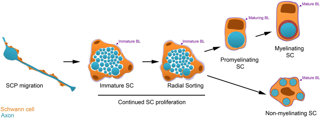
In the peripheral nervous system, Schwann cells are glial cells that are in intimate contact with axons throughout development. Schwann cells generate the insulating myelin sheath and provide vital trophic support to the neurons that they ensheathe. Schwann cell precursors arise from neural crest progenitor cells, and a highly ordered developmental sequence controls the progression of these cells to become mature myelinating or nonmyelinating Schwann cells. Here, we discuss both seminal discoveries and recent advances in our understanding of the molecular mechanisms that drive Schwann cell development and myelination with a focus on cell-cell and cell-matrix signaling events.

Keywords: Remak Schwann cell; Schwann cell; Schwann cell precursor; immature Schwann cell; myelinating Schwann cell; neural crest; peripheral nervous system; radial sorting.


Figures

Figure 1
Figure 1. Development of Myelinating and Non-myelinating Schwann Cells
Cartoon depicting Schwann cell (SC) development. SCs are orange and axons are blue. From left, SC precursors (SCPs), depicted longitudinally, are proliferative and migrate with growing axons. The remaining stages are depicted in cross-section. SCPs become immature SCs, which are associated with many axons. Immature SCs have ceased migration, remain proliferative, and form an immature basal lamina (BL, purple). The BL persists and matures through subsequent stages of SC development. During radial sorting, immature SCs interdigitate their cytoplasmic processes into the axon bundle, and promyelinating SCs are associated with a single axonal segment. Myelinating SCs spiral their membrane many times around the axonal segment to form the myelin sheath. Immature SCs can also develop into Non-myelinating SCs. A Remak SC, entheathing multiple small caliber axons is depicted.
Figure 2
Figure 2. Axo-glial interactions during radial sorting and myelination: novel concepts
The image depicts molecules that were discovered recently and mediate signaling between Schwann cells and axons, or Schwann cells and the ECM, during radial sorting and myelination.
Figure 3
Figure 3. ECM Molecules and GPCRs in SC Myelination – Recent Advances
From left, clockwise. In the SC basal lamina, Laminin 211 binds traditional (Integrins αββ1 and α7β1, dystroglycan) and unconventional (GPR126) receptors. Conversely, multiple ECM constituents bind the same receptor: integrin α6β1 can bind Laminin 211 and 411; The N-terminal fragment (NTF) of GPR126 can bind Laminin 211 and Col IV. The NTF must be modulated, perhaps by physical removal, in order to expose the Stachel tethered agonist (S) to activate the C-terminal fragment (CTF) of GPR126. CTF activation promotes cAMP accumulation, which activates PKA. This likely activates the cAMP-response element binding protein (CREB) pathway and phosphorylates NFkB, and a major downstream effector of cAMP in SCs is Oct-6. NRG1 type III intramembrane cleavage induces the expression of L-PGDS, which is released to produce prostaglandin D2 (PGD2). PGD2 then binds to GPR44 expressed on SCs membrane. Activation of GPR44 induces NFATc4 dephosphorylation and nuclear translocation to promote myelination.

References

    1. Aguayo AJ, Epps J, Charron L, Bray GM. Multipotentiality of Schwann cells in cross-anastomosed and grafted myelinated and unmyelinated nerves: quantitative microscopy and radioautography. Brain Res. 1976;104:1–20. - PubMed
    1. Aguayo AJ, Peyronnard JM, Bray GM. A quantitative ultrastructural study of regeneration from isolated proximal stumps of transected unmyelinated nerves. J Neuropath Experim Neurol. 1973;32:256–270. - PubMed
    1. Anliker B, Choi JW, Lin ME, Gardell SE, Rivera RR, Kennedy G, Chun J. Lysophosphatidic acid (LPA) and its receptor, LPA1, influence embryonic schwann cell migration, myelination, and cell-to-axon segregation. Glia. 2013;61:2009–2022. - PMC - PubMed
    1. Arroyo EJ, Bermingham JR, Jr, Rosenfeld MG, Scherer SS. Promyelinating Schwann cells express Tst-1/SCIP/Oct-6. J Neurosci. 1998;18:7891–7902. - PMC - PubMed
    1. Arter J, Wegner M. Transcription factors Sox10 and Sox2 functionally interact with positive transcription elongation factor b in Schwann cells. J Neurochem. 2015;13:384–393. - PubMed

Publication types