. 2015 Jun 5;290(23):14692-704.
doi: 10.1074/jbc.M114.633461. Epub 2015 Apr 28.

p47phox-Nox2-dependent ROS Signaling Inhibits Early Bone Development in Mice but Protects against Skeletal Aging

Affiliations

p47phox-Nox2-dependent ROS Signaling Inhibits Early Bone Development in Mice but Protects against Skeletal Aging

Jin-Ran Chen et al. J Biol Chem. .

Abstract

Bone remodeling is age-dependently regulated and changes dramatically during the course of development. Progressive accumulation of reactive oxygen species (ROS) has been suspected to be the leading cause of many inflammatory and degenerative diseases, as well as an important factor underlying many effects of aging. In contrast, how reduced ROS signaling regulates inflammation and remodeling in bone remains unknown. Here, we utilized a p47(phox) knock-out mouse model, in which an essential cytosolic co-activator of Nox2 is lost, to characterize bone metabolism at 6 weeks and 2 years of age. Compared with their age-matched wild type controls, loss of Nox2 function in p47(phox-/-) mice resulted in age-related switch of bone mass and strength. Differences in bone mass were associated with increased bone formation in 6-week-old p47(phox-/-) mice but decreased in 2-year-old p47(phox-/-) mice. Despite decreases in ROS generation in bone marrow cells and p47(phox)-Nox2 signaling in osteoblastic cells, 2-year-old p47(phox-/-) mice showed increased senescence-associated secretory phenotype in bone compared with their wild type controls. These in vivo findings were mechanistically recapitulated in ex vivo cell culture of primary fetal calvarial cells from p47(phox-/-) mice. These cells showed accelerated cell senescence pathway accompanied by increased inflammation. These data indicate that the observed age-related switch of bone mass in p47(phox)-deficient mice occurs through an increased inflammatory milieu in bone and that p47(phox)-Nox2-dependent physiological ROS signaling suppresses inflammation in aging.

Keywords: TNFα; aging; osteoblast; reactive oxygen species.


Figures

FIGURE 1.
FIGURE 1.
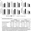
Increased bone quantity and quality in 6-week-old p47phox−/− mice but reversed in 2-year-old p47phox−/− mice compared with their age-matched respective controls. A, quantitative pQCT analysis of the proximal tibial in 6-week-old and 2-year-old p47phox−/− mice versus their respective wild type controls (n = 8 mice). Trabecular BMD (mg/cm3), total BMD, cortical BMD, and total bone mineral content (BMC) are presented. B, femur three-point bending test to determine bone strength was performed; parameters of peak load and stiffness are presented. C, bone formation and resorption markers in serum were measured. The data are expressed as means ± S.E. (n = 8 per group). * > wild type; # < wild type p < 0.05.
FIGURE 2.
FIGURE 2.
Bone histomorphometric and microcomputed tomography analysis. A and B, 6-week-old (A) and 2-year-old (B) bone histomorphometric parameters from p47phox−/− and control mice. The pictures on the left are representatives of histomorphometric Masson staining from p47phox−/− and control mice with different ages. Numbers and bar graphs are from histomorphometric reading. Ob. No, osteoblast number; Oc. No, osteoclast number; ES/BS, bone erosion surface per bone surface (%). C, bar graphs show quantitative microcomputed tomography analysis of the proximal tibial in p47phox−/− and wild type control mice (n = 5 per group). BV/TV, bone volume/tissue volume (%); J (mm4), polar moment of inertia. The data are expressed as means ± S.E. (n = 5 per group). Significant differences (p < 0.05) are indicated by * and # compared with control animals.
FIGURE 3.
FIGURE 3.
Nox2 signaling is not required for osteoblast differentiation. A, ROS measurements in bone marrow cells from p47phox−/− and wild type control mice using flow cytometrics. Three animal samples per group were presented, bone marrow cells were loaded with 10 m 2,7-DCF-DA for 30 min, living cells were collected, and ROS were monitored (2,7-DCF-DA fluorescence) by flow cytometry. The numbers in the upper right corners indicate cell number in this panel with ROS positive. B and C, basal (B) and phorbol 12-myristate 13-acetate (PMA)-stimulated (C) H2O2 generation was measured in wild type and p47phox−/− mouse neonatal calvarial cell cultures using the Amplex Red hydrogen peroxide/peroxidase assay kit. D, neonatal calvarial cells from wild type or p47phox−/− mice were cultured in 6-well plates for 12 days, and ALP staining was performed. E, Nox4 shRNA was transfected into calvarial cells from either wild type or p47phox−/− mice. Nox4 expression in these transfected cells were determined using Western blots. F, after 24 h, ALP gene expression was examined in calvarial cells from either wild type or p47phox−/− mice with or without Nox4 shRNA using real time PCR. Significant differences (p < 0.05) are indicated by * and # compared with either wild type mice or control.
FIGURE 4.
FIGURE 4.
Increased inflammation in bone from 2-year-old p47phox−/− mice. A, proteins were isolated from femur and Western blots showing expression of RANKL, TNFα and MMP9, and Col-1 (collagen type 1) and Runx2 in 6-week-old and 2-year-old p47phox−/− mice compared with those from wild type mice. B, decalcified vertebrae section antibody immunostaining for TNFα (stained red). C, real time PCR for the expression of TNFα, MMP9, ALP, osteocalcin (OC), and Runx2 in bone from 6-week-old and 2-year-old wild type and p47phox−/− mice. D, heat map analysis of mouse inflammation antibody array C1 using proteins isolated from mouse vertebrae. Three pooled samples per group. Significant differences (p < 0.05) are indicated by * and # compared with respective wild type and p47phox−/− mice.
FIGURE 5.
FIGURE 5.
Increased senescence pathway in bone from 2-year-old p47phox−/− mice. A, proteins were isolated from bone from 6-week-old and 2-year-old wild type and p47phox−/− mice, and SABG activity was measured (see “Experimental Procedures”). B, RNA was isolated from bone from 6-week-old and 2-year-old wild type and p47phox−/− mice; real time PCR was performed for p16 and p53 gene expression. C, real time PCR was performed for cyclin A and B gene expression. Significant differences (p < 0.05) are indicated by * compared with respective wild type control mice.
FIGURE 6.
FIGURE 6.
Passage 8 neonatal calvarial cells from p47phox−/− mice are senescent. Neonatal calvarial cells were isolated from either wild type or p47phox−/− mice and were passaged into new 6-well plates after cells reached 80% confluent, and cell passaging was continued 20 times. A, at passage 8, calvarial cells from p47phox−/− mice significantly decreased their proliferation (left, proliferation assay) and differentiation (right, ALP staining) compared with calvarial cells from wild type control mice. B, RNA was collected from calvarial cells at passages 2 and 8; real time PCR showed that passage 8 calvarial cells from p47phox−/− mice decreased osteoblastic cell differentiation markers ALP and Runx2 but increased cell senescent markers p16 and p53 compared with cells from wild type control mice. C, SABG activity measurement from collected protein samples showed similar changes to p16 and p53 gene expression. D, cell passage was continued until 20, SABG blue staining showed remarkable changes in cells from p47phox−/− mice. E, Western blot for TNFα from protein isolated from passage 8 and 20 calvarial cells from either wild type or p47phox−/− mice. F, heat map analysis of mouse inflammation antibody array C1 using proteins isolated from passage 20 cells in triplicate. Significant differences (p < 0.05) are indicated by * compared with respective cells from wild type control mice.
FIGURE 7.
FIGURE 7.
Transient versus sustained inhibition of Nox activity resulted in different effects on osteoblastic cell senescence signaling and inflammation. A, RNA was collected from calvarial cells at passages 2, 8, and 20 from p47phox−/− and control mice; real time PCR showed increased expression of cyclin A and B in passage 2 cells from p47phox−/− but decreased expression of both cyclin A and B in passage 8 and 20 cells from p47phox−/− compared with cells from control. B, calvarial cells from control mice were treated with Nox pan inhibitor DPI for 4, 24, and 72 h with three different doses. Proteins were collected for SABG activity measurement. C, calvarial cells from control mice were treated with Nox pan inhibitor DPI 50 nm for 4, 24, and 72 h, and RNA were collected for TNFα mRNA expression using real time PCR. Significant differences (p < 0.05) are indicated by * compared with respective cells from wild type control mice or vehicle-treated cells.

References

    1. Kassem M., Marie P. J. (2011) Senescence-associated intrinsic mechanisms of osteoblast dysfunctions. Aging Cell 10, 191–197 - PubMed
    1. Almeida M. (2012) Aging mechanisms in bone. Bonekey Rep. 1, 102. - PMC - PubMed
    1. Tsukagoshi H., Busch W., Benfey P. N. (2010) Transcriptional regulation of ROS controls transition from proliferation to differentiation in the root. Cell 143, 606–616 - PubMed
    1. Bedard K., Krause K. H. (2007) The NOX family of ROS-generating NADPH oxidases: physiology and pathophysiology. Physiol. Rev. 87, 245–313 - PubMed
    1. Hagenow K., Gelderman K. A., Hultqvist M., Merky P., Bäcklund J., Frey O., Kamradt T., Holmdahl R. (2009) Ncf1-associated reduced oxidative burst promotes IL-33R+ T cell-mediated adjuvant-free arthritis in mice. J. Immunol. 183, 874–881 - PubMed

Publication types

MeSH terms