Review
. 2015 May 1;142(9):1561-71.
doi: 10.1242/dev.114926.

Sensory hair cell development and regeneration: similarities and differences

Affiliations
Review

Sensory hair cell development and regeneration: similarities and differences

Patrick J Atkinson et al. Development. .

Abstract

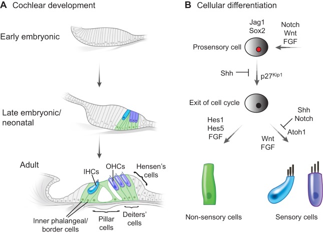
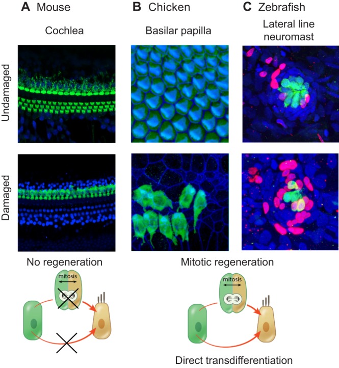
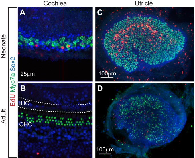
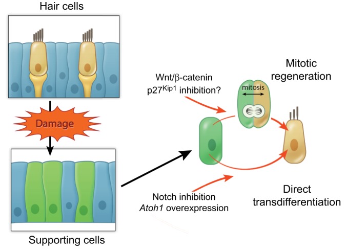
Sensory hair cells are mechanoreceptors of the auditory and vestibular systems and are crucial for hearing and balance. In adult mammals, auditory hair cells are unable to regenerate, and damage to these cells results in permanent hearing loss. By contrast, hair cells in the chick cochlea and the zebrafish lateral line are able to regenerate, prompting studies into the signaling pathways, morphogen gradients and transcription factors that regulate hair cell development and regeneration in various species. Here, we review these findings and discuss how various signaling pathways and factors function to modulate sensory hair cell development and regeneration. By comparing and contrasting development and regeneration, we also highlight the utility and limitations of using defined developmental cues to drive mammalian hair cell regeneration.

Keywords: Atoh1; FGF; Notch; Shh; Wnt; p27Kip1, Cdkn1b.


Figures

Fig. 1.
Fig. 1.
Cell fate decisions and the development of the mammalian organ of Corti. (A) An overview of cochlear development, highlighting the location of inner hair cells (IHCs, blue), outer hair cells (OHCs, purple) and various supporting cell subtypes (green). (B) A simplified schematic highlighting some of the signaling pathways and factors responsible for the specification of prosensory, sensory and non-sensory cells. The FGF, Notch and Wnt signaling pathways, together with the transcription factor Sox2, are required for prosensory cell specification and/or maintenance. The timing of terminal mitosis and the upregulation of the cell cycle inhibitor p27Kip1 in Sox2-positive prosensory cells are governed by Shh signaling. Wnt and FGF signaling are required for Atoh1 expression and the induction of sensory cell fate, which is restricted by the Notch and Shh pathways. By contrast, the activation of Notch target genes (Hes1 and Hes5) promotes a supporting cell fate, with FGF signals regulating differentiation into supporting cell subtypes.
Fig. 2.
Fig. 2.
Regenerative capacities of the adult mouse cochlea, the chicken basilar papilla and the zebrafish lateral line system. (A) No sensory hair cell regeneration occurs in the adult mouse cochlea. Shown are the basal turns of postnatal day (P) 28-30 cochleae from control (undamaged) mice and from mice that were treated with sisomicin and furosemide at P21. Immunostaining was performed to detect myosin VIIa (green) and Sox2 (blue); the reduction in myosin VIIa-labeled hair cells indicates that the damaged hair cells are not able to regenerate. (B) Hair cell regeneration in the chicken basilar papilla. Organs were harvested from control chicks and from those that had received daily injections of streptomycin in the previous week. Myosin VIIa-labeled hair cells are in green and phalloidin staining (F-actin) is in blue. Image courtesy of M. Warchol. (C) Hair cell regeneration in the zebrafish lateral line system. Images show posterior lateral line neuromasts from control zebrafish and from those that were damaged with neomycin. Hair cells are labeled by GFP in green, while BrdU (red) marks proliferating cells and DAPI staining (blue) marks nuclei. Regeneration in the neuromast populations, as indicated by the number of BrdU-positive cells, occurs primarily through mitotic regeneration. Image courtesy of T. Piotrowski and A. Romero-Carvajal.
Fig. 3.
Fig. 3.
Comparison of the modes and capacity of hair cell regeneration in the neonatal and adult mouse cochlea and utricle. (A) Limited mitotic hair cell regeneration occurs in the neonatal cochlea following diphtheria toxin-induced damage. Shown is the apical turn of the cochlea from P7 Pou4f3DTR/+ mice, which received diphtheria toxin at P1 to ablate hair cells. Immunostaining for myosin VIIa (green) highlights hair cells, while Sox2-positive supporting cells are in blue. The presence of hair cells labeled with EdU (red) indicates mitotic hair cell regeneration. Image courtesy of R. Chai. (B) Conversely, no mitotic response (EdU labeling) or hair cell regeneration occurs in the damaged adult cochlea. The image shows a cochlea from Pou4f3DTR/+ mice that received diphtheria toxin one week previously. There is a complete loss of inner hair cells (IHC) and a partial loss of outer hair cells (OHC; green). (C) By contrast, robust mitotic hair cell regeneration (as indicated by the abundance of EdU-labeled hair cells) is observed in the neonatal utricle. The image is of a utricle from P30 Pou4f3DTR/+ mice that received diphtheria toxin at P1 to ablate hair cells. Image courtesy of R. Chai and T. Wang. (D) Scant proliferation and regeneration are observed in the damaged mature utricle. Shown is a utricle from an adult mouse previously treated with a vestibulotoxin.
Fig. 4.
Fig. 4.
Proposed mechanisms of hair cell regeneration. Supporting cells can classically regenerate hair cells via two mechanisms: mitotic hair cell regeneration and direct transdifferentiation. Enhancing the Wnt/β-catenin signaling pathway or inhibiting cell cycle inhibitors such as p27Kip1 may constitute approaches to enhance mitotic regeneration. By contrast, Notch inhibition and Atoh1 overexpression may promote the direct conversion of supporting cells into hair cells.

References

    1. Ahmed M., Wong E. Y. M., Sun J., Xu J., Wang F. and Xu P.-X. (2012). Eya1-Six1 interaction is sufficient to induce hair cell fate in the cochlea by activating Atoh1 expression in cooperation with Sox2. Dev. Cell 22, 377-390 10.1016/j.devcel.2011.12.006 - DOI - PMC - PubMed
    1. Alvarado D. M., Hawkins R. D., Bashiardes S., Veile R. A., Ku Y.-C., Powder K. E., Spriggs M. K., Speck J. D., Warchol M. E. and Lovett M. (2011). An RNA interference-based screen of transcription factor genes identifies pathways necessary for sensory regeneration in the avian inner ear. J. Neurosci. 31, 4535-4543 10.1523/JNEUROSCI.5456-10.2011 - DOI - PMC - PubMed
    1. Atkinson P. J., Wise A. K., Flynn B. O., Nayagam B. A. and Richardson R. T. (2014). Hair cell regeneration after ATOH1 gene therapy in the cochlea of profoundly deaf adult guinea pigs. PLoS ONE 9, e102077 10.1371/journal.pone.0102077 - DOI - PMC - PubMed
    1. Barker N. and Clevers H. (2006). Mining the Wnt pathway for cancer therapeutics. Nat. Rev. Drug Discov. 5, 997-1014 10.1038/nrd2154 - DOI - PubMed
    1. Barker N., van Es J. H., Kuipers J., Kujala P., van den Born M., Cozijnsen M., Haegebarth A., Korving J., Begthel H., Peters P. J. et al. (2007). Identification of stem cells in small intestine and colon by marker gene Lgr5. Nature 449, 1003-1007 10.1038/nature06196 - DOI - PubMed

Publication types

MeSH terms

Substances