. 2015 Apr 29;10(4):e0124410.
doi: 10.1371/journal.pone.0124410. eCollection 2015.

MicroRNA-302a Suppresses Tumor Cell Proliferation by Inhibiting AKT in Prostate Cancer

Affiliations

MicroRNA-302a Suppresses Tumor Cell Proliferation by Inhibiting AKT in Prostate Cancer

Gui-Ming Zhang et al. PLoS One. .

Erratum in

Abstract

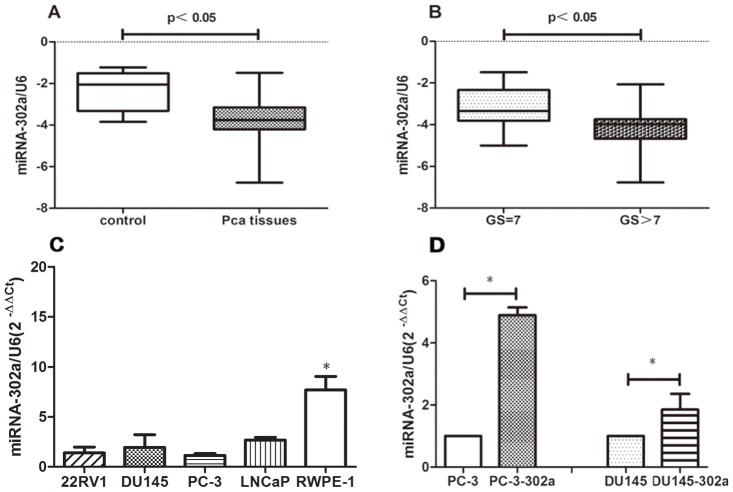
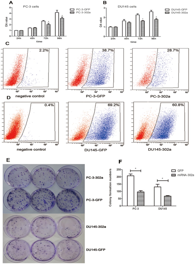
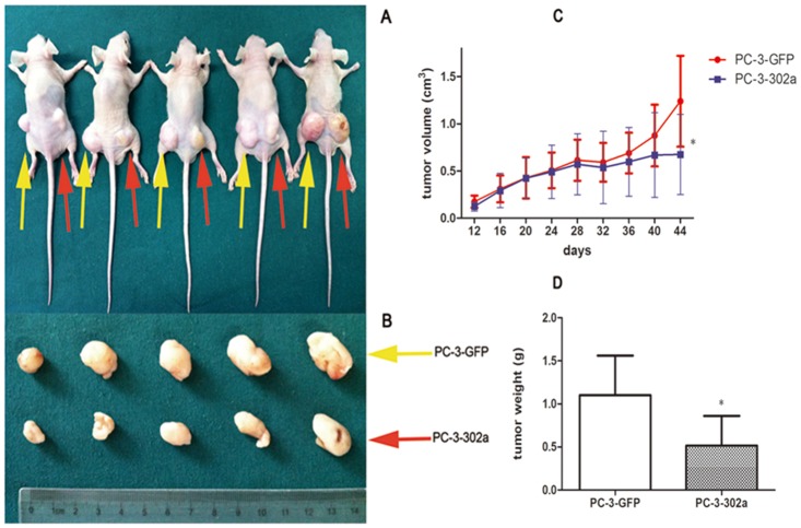
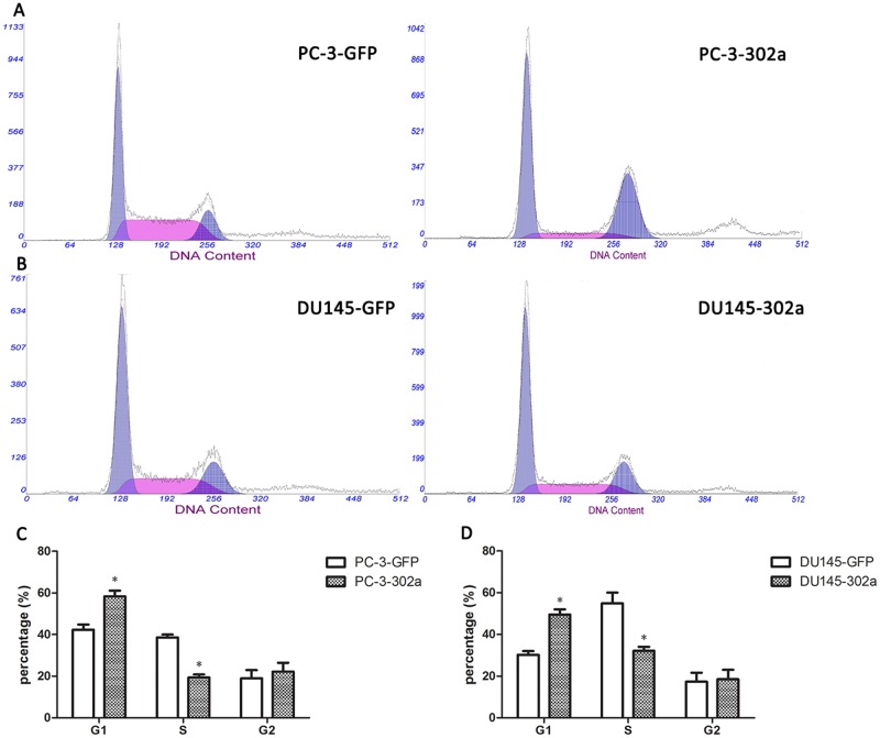
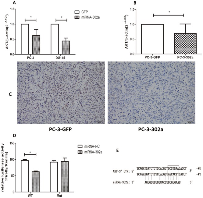
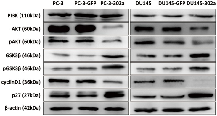
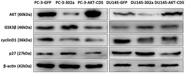
Micro (mi) RNAs are important regulators involved in various physical and pathological processes, including cancer. The miRNA-302 family has been documented as playing a critical role in carcinogenesis. In this study, we investigated the role of miRNA-302a in prostate cancer (PCa). MiRNA-302a expression was detected in 44 PCa tissues and 10 normal prostate tissues, and their clinicopathological significance was analyzed. Cell proliferation and cell cycle analysis were performed on PCa cells that stably expressed miRNA-302a. The target gene of miRNA-302a and the downstream pathway were further investigated. Compared with normal prostate tissues, miRNA-302a expression was downregulated in PCa tissues, and was even lower in PCa tissues with a Gleason score ≥8. Overexpression of miRNA-302a induced G1/S cell cycle arrest in PCa cells, and suppressed PCa cell proliferation both in vitro and in vivo. Furthermore, miRNA-302a inhibits AKT expression by directly binding to its 3΄ untranslated region, resulting in subsequent alterations of the AKT-GSK3β-cyclin D1 and AKT-p27Kip1 pathway. These results reveal miRNA-302a as a tumor suppressor in PCa, suggesting that miRNA-302a may be used as a potential target for therapeutic intervention in PCa.


Conflict of interest statement

Competing Interests: The authors have declared that no competing interests exist.

Figures

Fig 1
Fig 1. MiRNA-302a expression profiles in PCa tissue, normal prostate tissue, PCa cells and normal prostate epithelial cells.
(A) MiRNA-302a expression was downregulated in PCa tissue compared with normal prostate tissue. (B) MiRNA-302a expression was lower in PCa tissues with GS > 7 compared with those with GS = 7. (C) Lower levels of miRNA-302a expression were detected in four human PCa cell lines compared with normal prostate epithelial cells. (D) High levels of miRNA-302a expression were detected in PCa cells stably expressing miRNA-302a (*P<0.05).
Fig 2
Fig 2. Overexpression of miRNA-302a significantly inhibits cell proliferation in PCa cells in vitro.
A CCK-8 assay was performed to measure proliferation in (A) PC-3 and (B) DU145 cells. Data represent the mean ± standard deviation of the optical density (OD) value detected at 450 nm from three independent experiments. Cell proliferation was detected in (C) PC-3 and (D) DU145 cells using EdU assay analyzed by flow cytometry. (E, F) Colony formation assays indicated fewer colonies in miRNA-302a overexpressing PCa cells. (*P<0.05).
Fig 3
Fig 3. Overexpression of miRNA-302a significantly inhibits cell proliferation in PCa cells in vivo.
PC-3-GFP cells and PC-3-302a cells were injected into the left and right posterior flank of BALB/c nude mice, respectively (A, B). The tumor volume (C) and mass (D) in the PC-3-302a group were notably lower than in the PC-3-GFP group. (* P<0.05).
Fig 4
Fig 4. Overexpression of miRNA-302a induces G1/S phase cell cycle arrest in PCa cells.
The proportion of cells in G1-phase increased significantly in PC-3-302a cells (A, C) and DU145-302a cells (B, D) compared with controls, while the proportion of cells in the S phase were notably less than the controls. (* P<0.05).
Fig 5
Fig 5. MiRNA-302a suppresses AKT expression by directly targeting its 3΄ untranslated region.
AKT mRNA expression was remarkably suppressed in (A) PC-3-302a and DU145-302a cells, and (B) PC-3-302a tumors (five independent tumors were detected). (C) Immunohistochemistry staining indicated lower expression of AKT in PC-3-302a tumors. (D) Relative luciferase activity was notably suppressed in wild-type AKT-3΄ untranslated region (UTR) transfected cells. (E) Schematic representation of the luciferase reporter, which carried the wild-type or mutant AKT-3’ UTR. (* P<0.05).
Fig 6
Fig 6. Overexpression of miRNA-302a in PCa cells triggers alterations in the AKT-GSK3β-cyclin D1 and AKT-p27 Kip1signaling pathways.
Western blot analyses showed downregulated AKT, phosphorylated AKT (pAKT) and cyclin D1 levels, and upregulated GSK3β,pGSK3β and p27Kip1 levels in miRNA-302a overexpressing PCa cells. PI3K levels were not affected.
Fig 7
Fig 7. Alterations of AKT-GSK3β-cyclin D1 and AKT-p27 Kip1 signaling pathways after AKT restoration in miRNA-302a overexpressed PCa cells.
Western blot analyses showed rescued expression of AKT, and inhibited expression of GSK3β and p27Kip1 levels by AKT restoration. However, cyclin D1 levels were not rescued.

References

    1. Jemal A, Bray F, Center MM, Ferlay J, Ward E, Forman D (2011) Global cancer statistics. CA Cancer J Clin 61: 69–90. 10.3322/caac.20107 - DOI - PubMed
    1. He J, Chen WQ (2012) Chinese cancer registry annual report. Military Medical Science Press, Beijing.
    1. Coleman RE (2006) Clinical features of metastatic bone disease and risk of skeletal morbidity. Clin Cancer Res 12: 6243s–6249s. - PubMed
    1. Ambros V (2004) The functions of animal microRNAs. Nature 431: 350–355. - PubMed
    1. Bartel DP (2004) MicroRNAs: genomics, biogenesis, mechanism, and function. Cell 116: 281–297. - PubMed

Publication types

MeSH terms