. 2015 Apr 29:5:9841.
doi: 10.1038/srep09841.

SIRT7 inactivation reverses metastatic phenotypes in epithelial and mesenchymal tumors

Affiliations

SIRT7 inactivation reverses metastatic phenotypes in epithelial and mesenchymal tumors

Shivani Malik et al. Sci Rep. .

Abstract

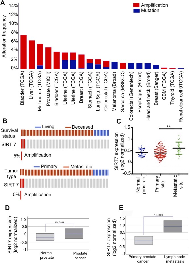
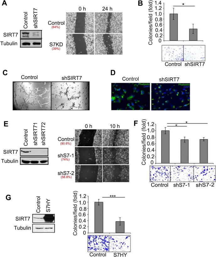
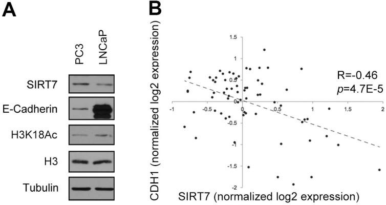
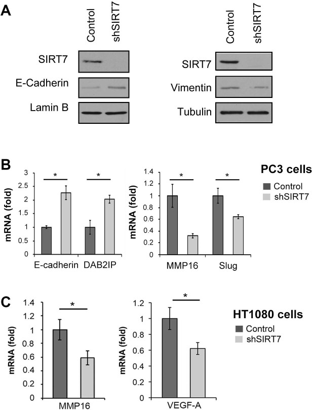
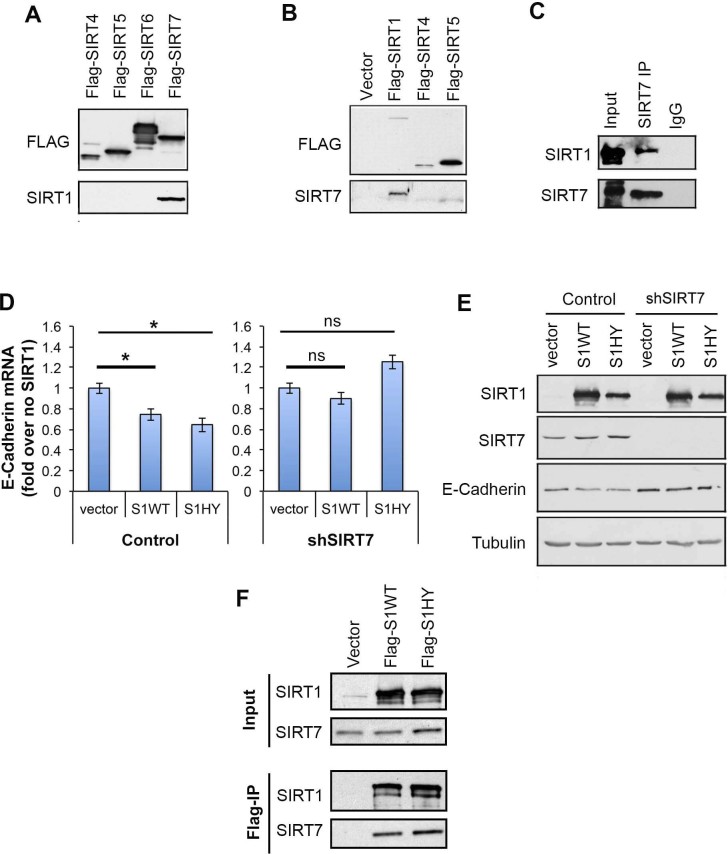
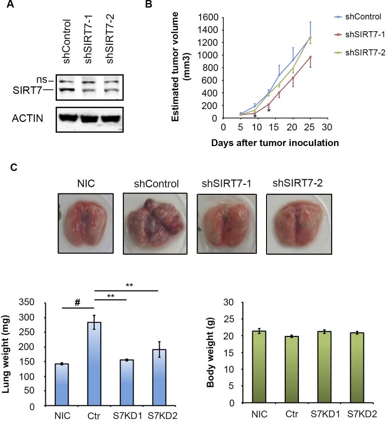
Metastasis is responsible for over 90% of cancer-associated mortality. In epithelial carcinomas, a key process in metastatic progression is the epigenetic reprogramming of an epithelial-to-mesenchymal transition-like (EMT) change towards invasive cellular phenotypes. In non-epithelial cancers, different mechanisms must underlie metastatic change, but relatively little is known about the factors involved. Here, we identify the chromatin regulatory Sirtuin factor SIRT7 as a key regulator of metastatic phenotypes in both epithelial and mesenchymal cancer cells. In epithelial prostate carcinomas, high SIRT7 levels are associated with aggressive cancer phenotypes, metastatic disease, and poor patient prognosis, and depletion of SIRT7 can reprogram these cells to a less aggressive phenotype. Interestingly, SIRT7 is also important for maintaining the invasiveness and metastatic potential of non-epithelial sarcoma cells. Moreover, SIRT7 inactivation dramatically suppresses cancer cell metastasis in vivo, independent of changes in primary tumor growth. Mechanistically, we also uncover a novel link between SIRT7 and its family member SIRT1, providing the first demonstration of direct interaction and functional interplay between two mammalian sirtuins. Together with previous work, our findings highlight the broad role of SIRT7 in maintaining the metastatic cellular phenotype in diverse cancers.


Conflict of interest statement

Dr. Chua’s research is partly funded by Daiichi Sankyo company. Dr. Michishita-Kioi, Dr. Tanaka, and Dr. Aonuma are employees of Daiichi Sankyo Group Companies.

Figures

Figure 1
Figure 1. SIRT7 amplification and over-expression is associated with metastatic disease and poor prognosis.
(A), Analysis of several cBio Cancer Genomics datasets shows that SIRT7 is amplified in many epithelial cancers and in mesenchymal sarcomas. (B), Analysis of a reported mutational landscape of metastatic prostate cancer shows exclusive amplification of SIRT7 in prostate cancer patients with metastatic disease and poor survival. (C), Meta-analysis of the prostate cancer dataset using the Oncomine database showing significantly higher expression of SIRT7 in metastatic tumor sites compared to primary tumor or normal prostate tissues. (D), SIRT7 expression in prostate cancer patient tissues versus normal prostate controls. (E), SIRT7 expression in primary prostate cancer tumors versus metastatic tumor tissue. In (D), and (E), levels are log2 normalized values (median, lower and upper quartiles are shown), retrieved from Lapointe et al.
Figure 2
Figure 2. SIRT7 promotes cancer cell migration and invasiveness.
(A), Wound healing assay showing reduced migration of SIRT7-depleted (shSIRT7) PC3 cells. (B), Transwell assay showing reduced invasion of SIRT7 depleted PC3 cells. (C), 3D Matrigel assay showing reduced invasive growth of SIRT7 depleted cells through surrounding matrix. Images taken 3 days after plating of cells. (D), Phalloidin staining of F-actin revealed a more collapsed actin cytoskeleton in SIRT7-depleted PC3 cells compared to controls. (E), Impaired cell migration and wound healing in SIRT7- depleted HT1080 cells. (F), Impaired invasion of SIRT-depleted HT1080 cells in Transwell assay. (G), Reduced invasion of HT1080 cells overexpressing the catalytically inactive (S7-HY) SIRT7 protein in the Transwell assay.
Figure 3
Figure 3. SIRT7 expression correlates inversely with H3K18Ac and E-cadherin in prostate carcinoma.
(A), Western blot analysis showing enhanced expression of SIRT7 and lower levels of H3K18Ac and E-cadherin in PC3 cells as compared to LNCaP cells. (B), SIRT7 expression is negatively correlated with E-cadherin (CDH1) expression in human prostate cancers. cDNA microarray expression levels are log2 normalized values, retrieved from Lapointe et al. For CDH1, the average expression value from 4 cDNA probes was used. Correlation (R) and corresponding P-values are indicated. Western Blots were carried under same experimental conditions. Uncropped images are available in supplementary figure 1a.
Figure 4
Figure 4. SIRT7 regulates metastasis regulatory genes in epithelial and mesenchymal cancer cells.
(A), Western blot analysis showing decreased E-cadherin protein and increased Vimentin levels in SIRT7-deficient PC3 cells (shSIRT7). (B), RT-qPCR analysis showing modulation of E-cadherin, DAB2IP, MMP16, and Slug expression in SIRT7-depleted PC3. (C), RT-qPCR showing decreased expression of MMP16 and VEGF-A in SIRT7 depleted HT1080 cells. Western Blots were carried under same experimental conditions. Uncropped images are available in supplementary figure 1b.
Figure 5
Figure 5. SIRT7 physically interacts with SIRT1 and is required for SIRT1-mediated repression of E-cadherin.
(A), Western analysis showing co-immunoprecipitation (IP) of Flag-tagged SIRT7 and endogenous SIRT1. (B), Western analysis showing co-IP of Flag-tagged SIRT1 and endogenous SIRT7. (C), Western analysis showing co-IP of endogenous SIRT1 and SIRT7 proteins. (D), q-PCR analysis of the E-cadherin expression following overexpression of wild-type (S1WT) or catalytically inactive (S1HY) SIRT1 proteins in Control or SIRT7-depleted (shSIRT7) PC3 cells. (E), Western blot showing E-cadherin protein levels from cells used in D. (F), Western blot analysis showing co-immunoprecipitation of SIRT7 by the SIRT1 WT and HY mutant proteins. Western Blots were carried under same experimental conditions. Uncropped images are available in supplementary figure 1c.
Figure 6
Figure 6. SIRT7 depletion inhibits lung metastasis in vivo.
(A). Western blot of SIRT7 expression in cells with partial SIRT7 depletion. (B), Tumor volume of subcutaneous xenografts of the SIRT7-depleted and control cells. (C), Effects of SIRT7 depletion on lung metastasis following tail vein injection of immunodeficient mice with control (shcontrol) or SIRT7-depleted (shSIRT7-1, shSIRT7-2) cells. Non-Injected Control (NIC): normal lungs. *, ** indicate significant differences between the sh-control and shSIRT7 groups by Welch's T test (**: P < 0.005, *: P < 0.05). # indicates significant difference between the no injection control lungs and the sh-control HT1080-injected group (P < 0.005). ns, non-specific band.

References

    1. Paredes S., Villanova L. & Chua K. F. Molecular Pathways: Emerging Roles of Mammalian Sirtuin SIRT7 in Cancer. Clin Cancer Res: an official journal of the American Association for Cancer Research, 10.1158/1078-0432.CCR-13-1547 (2014). - DOI - PMC - PubMed
    1. Haigis M. C. & Sinclair D. A. Mammalian sirtuins: biological insights and disease relevance. Annu Rev Pathol 5, 253–295, 10.1146/annurev.pathol.4.110807.092250 (2010). - DOI - PMC - PubMed
    1. Barber M. F. et al. SIRT7 links H3K18 deacetylation to maintenance of oncogenic transformation. Nature 487, 114–118, 10.1038/nature11043 (2012). - DOI - PMC - PubMed
    1. Kim J. K. et al. Sirtuin7 oncogenic potential in human hepatocellular carcinoma and its regulation by the tumor suppressors MiR-125a-5p and MiR-125b. Hepatology, 10.1002/hep.26101 (2012). - DOI - PubMed
    1. Ashraf N. et al. Altered sirtuin expression is associated with node-positive breast cancer. Br J Cancer 95, 1056–1061 (2006). - PMC - PubMed

Publication types