. 2015 Apr 27;16(5):9406-19.
doi: 10.3390/ijms16059406.

Effects of mechanical stretching on the morphology and cytoskeleton of vaginal fibroblasts from women with pelvic organ prolapse

Affiliations

Effects of mechanical stretching on the morphology and cytoskeleton of vaginal fibroblasts from women with pelvic organ prolapse

Sumei Wang et al. Int J Mol Sci. .

Abstract

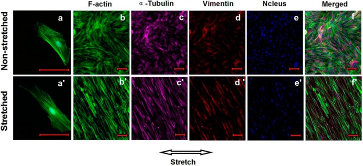
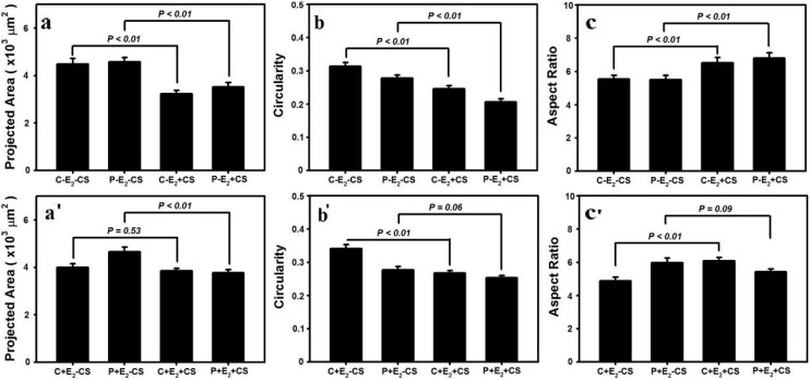
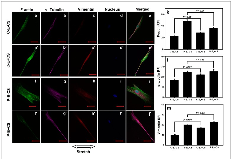
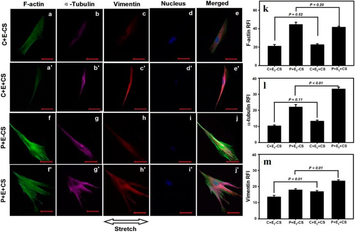
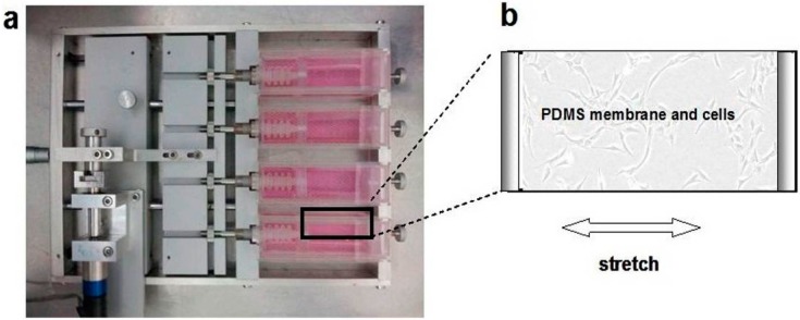
Mechanical load and postmenopausal hypoestrogen are risk factors for pelvic organ prolapse (POP). In this study, we applied a 0.1-Hz uniaxial cyclic mechanical stretching (CS) with 10% elongation and 10⁻⁸ M 17-β-estradiol to vaginal fibroblasts isolated from postmenopausal women with or without POP to investigate the effects of CS and estrogen on cell morphology and cytoskeletons of normal and POP fibroblasts. Under static culture condition, POP fibroblasts exhibited lower cell circularity and higher relative fluorescence intensities (RFIs) of F-actin, α-tubulin and vimentin. When cultured with CS, all fibroblasts grew perpendicular to the force and exhibited a decreased cell projection area, cell circularity and increased cell length/width ratio; normal fibroblasts exhibited increased RFIs of all three types of cytoskeleton, and POP fibroblasts exhibited a decreased RFI of F-actin and no significant differences of α-tubulin and vimentin. After being cultured with 17-β-estradiol and CS, normal fibroblasts no longer exhibited significant changes in the cell projection area and the RFIs of F-actin and α-tubulin; POP fibroblasts exhibited no significant changes in cell circularity, length/width ratio and F-actin even with the increased RFIs of α-tubulin and vimentin. These findings suggest that POP fibroblasts have greater sensitivity to and lower tolerance for mechanical stretching, and estrogen can improve the prognosis.


Figures

Figure 1
Figure 1
Morphological characteristics and immunocytochemical identification of vaginal fibroblasts (images acquired with an inverted microscope). Primary cultured pelvic organ prolapse (POP) fibroblasts (passage 0) after 72 h (a) and 7 days (b); POP fibroblasts at passages 4 (c) and 5 (d). Streptavidin-peroxidase (SP) immunohistochemical staining of POP fibroblasts (passage 4): The characteristic staining of vimentin (e), cytokeratin (f), α-smooth muscle actin (g), and negative control (h). Bar = 100 μm.
Figure 2
Figure 2
Effects of cyclic mechanical stretching (CS) on the orientations of cytoskeletons and vaginal fibroblasts. The static growth F-actin stress fibers (a) and fibroblasts (bf); The stretching growth F-actin stress fibers (a') and fibroblasts (b'–f'). Bar = 100 μm.
Figure 3
Figure 3
Effects of CS and 17-β-estradiol (E2) on the vaginal fibroblast projection area, circularity and length/width ratio (ac, a'–c'). Data are represented as the mean ± SE from triplicate trials.
Figure 4
Figure 4
Effects of CS on the cytoskeletons of vaginal fibroblasts. The comparisons in relative fluorescence intensities (RFIs) of the cytoskeletal proteins between the static-cultured fibroblasts and the CS-cultured fibroblasts (km). Images of the cytoskeletons (a'e' and ae, f'j' and fj). Bar = 50 μm. The RFIs of F-actin (k), α-tubulin (l), and vimentin (m) were measured and are represented as the mean ± SE from triplicate trials.
Figure 5
Figure 5
Effects of CS and E2 on vaginal fibroblast cytoskeletons. The comparisons in RFIs of the cytoskeletal proteins between the fibroblasts cultured in static conditions with E2 and cultured in the presence of CS concomitant E2 (km). Images of the cytoskeletons (a'e' and ae, f'j' and fj). Bar = 50 μm. The RFIs of F-actin (k), α-tubulin (l), and vimentin (m) were measured and are represented as the mean ± SE from triplicate trials.
Figure 6
Figure 6
The uniaxial CS was applied to the vaginal fibroblasts via the PDMS membrane fixed in-house of the cell stretching device (a); Vaginal fibroblasts sub-cultured on the PDMS membrane (b). Arrow indicates the stretching direction.

References

    1. Mokrzycki M.L., Mittal K., Smilen S.W., Blechman A.N., Porges R.F., Demopolous R.I. Estrogen and progesterone receptors in the uterosacral ligament. Obstet. Gynecol. 1997;90:402–404. doi: 10.1016/S0029-7844(97)00285-8. - DOI - PubMed
    1. Hendrix S.L., Clark A., Nygaard I., Aragaki A., Barnabei V., McTiernan A. Pelvic organ prolapse in the Women’s Health Initiative: Gravity and gravidity. Am. J. Obstet. Gynecol. 2002;186:1160–1166. doi: 10.1067/mob.2002.123819. - DOI - PubMed
    1. Olsen A.L., Smith V.J., Bergstrom J.O., Colling J.C., Clark A.L. Epidemiology of surgically managed pelvic organ prolapse and urinary incontinence. Obstet. Gynecol. 1997;89:501–506. doi: 10.1016/S0029-7844(97)00058-6. - DOI - PubMed
    1. Jelovsek J.E., Maher C., Barber M.D. Pelvic organ prolapse. Lancet. 2007;369:1027–1038. doi: 10.1016/S0140-6736(07)60462-0. - DOI - PubMed
    1. Kim C.M., Jeon M.J., Chung D.J., Kim S.K., Kim J.W., Bai S.W. Risk factors for pelvic organ prolapse. Int. J. Gynaecol. Obstet. 2007;98:248–251. doi: 10.1016/j.ijgo.2007.02.019. - DOI - PubMed

Publication types

LinkOut - more resources