. 2015 Apr 29;10(4):e0125396.
doi: 10.1371/journal.pone.0125396. eCollection 2015.

Potent Anti-HIV Chemokine Analogs Direct Post-Endocytic Sorting of CCR5

Affiliations

Potent Anti-HIV Chemokine Analogs Direct Post-Endocytic Sorting of CCR5

Claudia Bönsch et al. PLoS One. .

Abstract

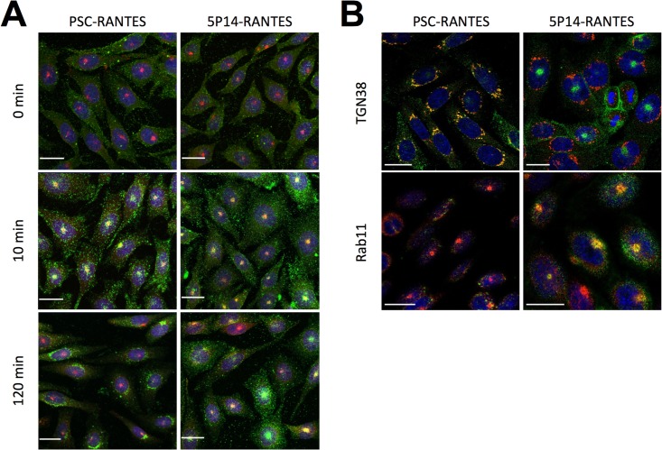
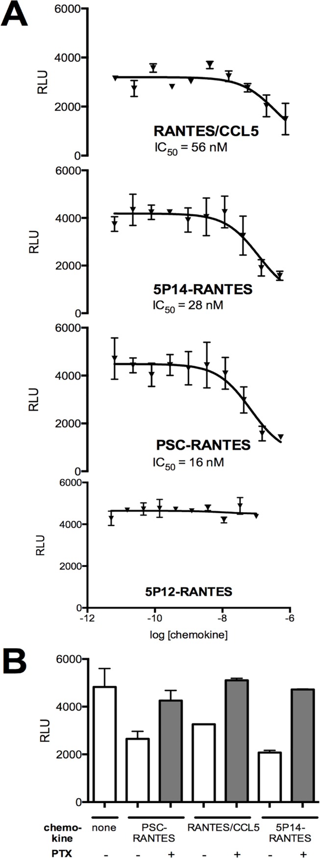
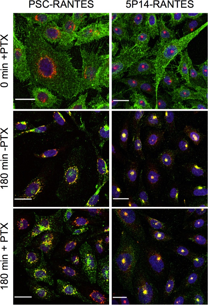
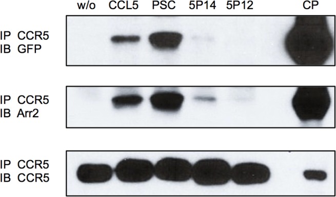
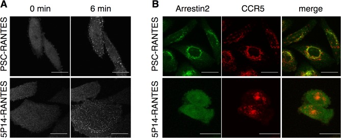
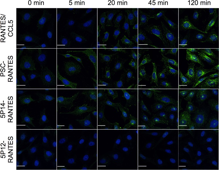
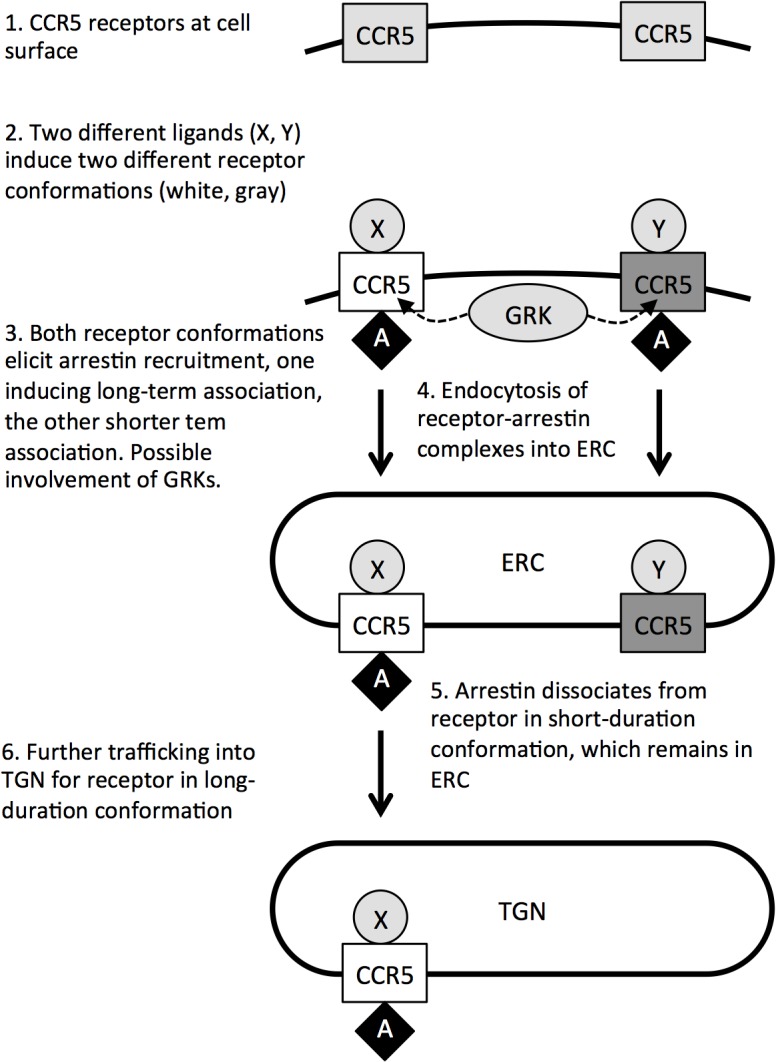
G protein-coupled receptors (GPCRs) are desensitized and internalized following activation. They are then subjected to post-endocytic sorting (degradation, slow recycling or fast recycling). The majority of research on post-endocytic sorting has focused on the role of sequence-encoded address structures on receptors. This study focuses on trafficking of CCR5, a GPCR chemokine receptor and the principal entry coreceptor for HIV. Using Chinese Hamster Ovary cells stably expressing CCR5 we show that two different anti-HIV chemokine analogs, PSC-RANTES and 5P14-RANTES, direct receptor trafficking into two distinct subcellular compartments: the trans-Golgi network and the endosome recycling compartment, respectively. Our results indicate that a likely mechanism for ligand-directed sorting of CCR5 involves capacity of the chemokine analogs to elicit the formation of durable complexes of CCR5 and arrestin2 (beta-arrestin-1), with PSC-RANTES eliciting durable association in contrast to 5P14-RANTES, which elicits only transient association.


Conflict of interest statement

Competing Interests: The authors have declared that no competing interests exist.

Figures

Fig 1
Fig 1. Ligand-directed post-endocytic sorting of CCR5.
A. CHO CCR5 cells growing on coverslips were pre-treated (60 min, 4°C) with 100 nM chemokines and anti-CCR5 antibody 3A9 (green), then washed and incubated at 37°C for the indicated times. Cells were then acid washed to remove cell surface antibody, then fixed, permeabilized, and labeled for the ERC marker Rab11 (red) and DAPI (blue, nuclear staining) prior to analysis by confocal scanning microscopy are shown, scale bar = 20 μm. Throughout the experiment the ERC marker (red) remains in a discrete supranuclear spot that is visble in the center of the nuclei (blue) in the maximum intensity projections. Initially, CCR5 (green) is localized at the cell surface, but after 10 min incubation with either ligand it translocates to colocalize with the ERC marker. After 120 min incubation, CCR5 on cells treated with PSC-RANTES subsequently relocates from the ERC to accumulate in a perinuclear site (ring-shaped staining around the nuclei), while CCR5 in cells treated with 5P14-RANTES remains colocalized with the ERC marker. B. Individual Z-slice images from an identical experiment in which cells incubated with the indicated chemokines for 180 min were also labeled for either the ERC marker, Rab11 or the TGN marker, TGN38 (red). Slices in which the marked compartment is most abundant (through the middle of the nucleus for TGN, just above the nucleus for ERC) were chosen. While in cells treated with PSC-RANTES for 180 min, CCR5 colocalizes with TGN38 and does not colocalize with Rab11, in cells treated with 5P14-RANTES for 180 CCR5 colocalizes with Rab11 and does not colocalize with TGN38 scale bar = 20 μm.
Fig 2
Fig 2. G protein signaling of chemokine analogs on CHO-CCR5 cells.
A. Gi/o protein signaling activity of chemokines at the indicated doses was determined by measuring reduction in forskolin-stimulated cAMP levels. Results shown (Relative Light Units, RLU) are the mean of duplicate readings, with error bars indicating the range. B. G protein signaling activity of chemokines (100 nM) was determined by measuring reduction in forskolin-stimulated cAMP levels. Where indicated CHO-CCR5 cells were pre-incubated (100 ng/mL, overnight) with Pertussis toxin (PTX). Results shown (Relative Light Units, RLU) are the mean of duplicate readings, with error bars indicating the range.
Fig 3
Fig 3. Ligand-directed post-endocytic sorting of CCR5 is not dependent on G protein signaling.
CHO CCR5 cells growing on glass coverslips were incubated overnight with Pertussis toxin (100 ng/mL), then washed and stimulated with 100 nM chemokines as indicated in the presence of anti-CCR5 antibody 3A9 (green) at 37°C. Cells were then acid washed (15 min 4°C) to remove cell surface antibody, fixed, permeabilized, and labelled for either the ERC marker Rab11 (5P14-RANTES) or the TGN marker TGN38 (PSC-RANTES) (red). After staining with DAPI nucleic acid stain (blue), cells were analysed by confocal microscopy. Maximum intensity projections are shown, scale bar = 20 μm. Throughout the experiment the ERC and TGN markers (red) remain either in a discrete supranuclear spot (ERC) or at a perinuclear site (ring-shaped staining around the nuclei, TGN). Initially, CCR5 (green) is localized at the cell surface, but after 10 min incubation with either ligand it translocates to the supranuclear spot (visibly colocalizing with the ERC marker used in the cells treated with 5P14-RANTES). After 120 min incubation, CCR5 on cells treated with PSC-RANTES subsequently relocates from the ERC to accumulate in a perinuclear site (ring-shaped staining around the nuclei, visibly colocalizing with the TGN marker), while CCR5 in cells treated with 5P14-RANTES remains colocalized with the ERC marker.
Fig 4
Fig 4. Ligand-elicited recruitment of arrestin2 to CCR5.
CHO-CCR5 cells stably transfected with arrestin2-GFP were treated with the indicated chemokines (100 nM, 50 min), washed extensively, crosslinked, washed again and lysed prior to immunoprecipitation with an anti-CCR5 antibody. Immunoprecipitates were subjected to Western blot using antibodies against GFP, arrestin2 and CCR5. CP; total cell pellet control, IP; antibody specificity used for immunoprecipitation, IB; antibody specificity used for immunoblot.
Fig 5
Fig 5. Spatial and temporal resolution of arrestin2-CCR5 association.
A CHO-CCR5 cells stably transfected with arrestin2-GFP were treated with chemokine analogs (100 nM) as indicated and the redistribution of arrestin2-GFP was followed by live fluorescence microscopy. Images captured prior to (0 min) and after ligand treatment (6 min) are shown. B CHO-CCR5 cells stably transfected with arrestin2-GFP (green), preincubated with rhodamine-labeled anti-CCR5 antibody (red), were washed and then incubated 90 min at 37°C with chemokine analogs (100 nM) prior to image capture. Maximal intensity projections are shown. Scale bar = 20 μm.
Fig 6
Fig 6. Ligand-induced GRK phosphorylation of CCR5.
CHO-CCR5 cells growing on coverslips were treated with the indicated chemokines (100 nM) for the indicated times, then washed, fixed, permeabilized and labeled with a monoclonal antibody specific for CCR5 phosphorylated on Serine 349 (green) and DAPI nucleic acid stain (blue). Maximal intensity projections are shown. Scale bar = 20 μm.
Fig 7
Fig 7. A model for ligand-directed post-endocytic sorting of CCR5.

References

    1. Fredriksson R, Lagerstrom MC, Lundin LG, Schioth HB. The G-protein-coupled receptors in the human genome form five main families. Phylogenetic analysis, paralogon groups, and fingerprints. Molecular pharmacology. 2003;63(6):1256–72. Epub 2003/05/23. 10.1124/mol.63.6.1256 . - DOI - PubMed
    1. Jacoby E, Bouhelal R, Gerspacher M, Seuwen K. The 7 TM G-protein-coupled receptor target family. ChemMedChem. 2006;1(8):761–82. Epub 2006/08/12. 10.1002/cmdc.200600134 . - DOI - PubMed
    1. Moore CA, Milano SK, Benovic JL. Regulation of receptor trafficking by GRKs and arrestins. Annual review of physiology. 2007;69:451–82. . - PubMed
    1. Shenoy SK, Lefkowitz RJ. beta-Arrestin-mediated receptor trafficking and signal transduction. Trends in pharmacological sciences. 2011;32(9):521–33. Epub 2011/06/18. 10.1016/j.tips.2011.05.002 - DOI - PMC - PubMed
    1. Hanyaloglu AC, Zastrow MV. Regulation of GPCRs by Endocytic Membrane Trafficking and Its Potential Implications. Annu Rev Pharmacol Toxicol. 2008;48:537–68. 10.1146/annurev.pharmtox.48.113006.094830 - DOI - PubMed

Publication types

MeSH terms

LinkOut - more resources