

Reversible and Asymptomatic Gyral and Subarachnoid Contrast Enhancement after Carotid Stenting
- PMID: 25923674
- PMCID: PMC4757145
- DOI: 10.1177/1971400915576630
Reversible and Asymptomatic Gyral and Subarachnoid Contrast Enhancement after Carotid Stenting
Abstract
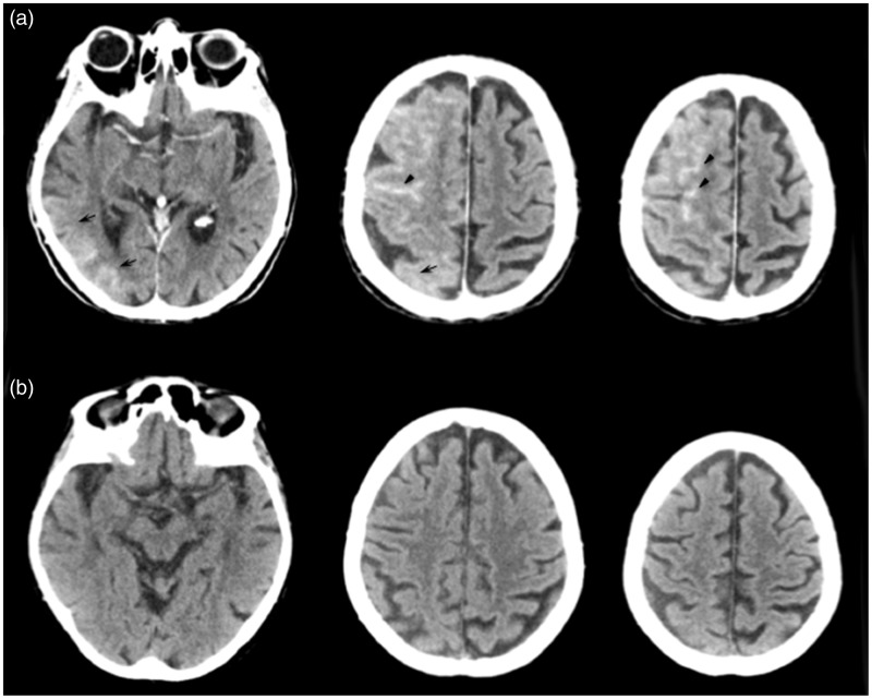
The presence of sulcal hyperdensity in patients after endovascular procedures is not necessarily attributable to hemorrhage. It may frequently indicate the absolute or concomitant extravasation of contrast material into the subarachnoid spaces.This case report describes the clinical case of an 84-year-old patient with 90% stenosis of the right internal carotid who presented with a diffuse gyral and sulcal hyperdensity in the right temporal-occipital and frontal lobes at routine post-carotid stenting (CAS) brain CT scan. The patient was asymptomatic and CT findings were interpreted as contrast enhancement hyperattenuation and no therapeutic decisions were made. A 24-hour follow-up brain CT demonstrated the complete resolution of the hyperdensity, confirming the diagnosis.In this patient we considered the concomitant presence of gyral and sulcal hyperdensity as the consequence of reversible damage to the blood-brain barrier (BBB) determining a transitory extravasation of contrast material. Asymptomatic gyral and subarachnoid contrast enhancement following CAS is generally indicative of benign and transitory damage to the BBB and is not to be misinterpreted as hemorrhage.
Keywords: carotid angioplasty and stenting; contrast extravasation.
© The Author(s) 2015 Reprints and permissions: sagepub.co.uk/journalsPermissions.nav.
Figures
References
-
- Kawabata Y, Fumihiko H, Miyake H, et al. Follow-up outcomes of self-expanding stents for carotid artery angioplasty at a single hospital. Neuroradiol J 2010; 23: 622–628. - PubMed
-
- Gado MH, Phelps ME, Coleman RE. An extravascular component of contrast enhancement in cranial computed tomography. Part I. The tissue-blood ratio of contrast enhancement. Radiology 1975; 117: 589–593. doi: 10.1148/117.3.589. - PubMed
-
- Eckel TS, Breiter SN, Monsein LH. Subarachnoid contrast enhancement after spinal angiography mimicking diffuse subarachnoid hemorrhage. Am J Roentgenol 1998; 170: 503–505. doi: 10.2214/ajr.170.2.9456974. - PubMed
Publication types
MeSH terms
Substances
LinkOut - more resources
Full Text Sources
Other Literature Sources
