ATP-Binding Cassette Transporter Structure Changes Detected by Intramolecular Fluorescence Energy Transfer for High-Throughput Screening
- PMID: 25924616
- PMCID: PMC4468642
- DOI: 10.1124/mol.114.096792
ATP-Binding Cassette Transporter Structure Changes Detected by Intramolecular Fluorescence Energy Transfer for High-Throughput Screening
Abstract
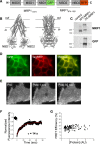
Multidrug resistance protein 1 (MRP1) actively transports a wide variety of drugs out of cells. To quantify MRP1 structural dynamics, we engineered a "two-color MRP1" construct by fusing green fluorescent protein (GFP) and TagRFP to MRP1 nucleotide-binding domains NBD1 and NBD2, respectively. The recombinant MRP1 protein expressed and trafficked normally to the plasma membrane. Two-color MRP1 transport activity was normal, as shown by vesicular transport of [(3)H]17β-estradiol-17-β-(D-glucuronide) and doxorubicin efflux in AAV-293 cells. We quantified fluorescence resonance energy transfer (FRET) from GFP to TagRFP as an index of NBD conformational changes. Our results show that ATP binding induces a large-amplitude conformational change that brings the NBDs into closer proximity. FRET was further increased by substrate in the presence of ATP but not by substrate alone. The data suggest that substrate binding is required to achieve a fully closed and compact structure. ATP analogs bind MRP1 with reduced apparent affinity, inducing a partially closed conformation. The results demonstrate the utility of the two-color MRP1 construct for investigating ATP-binding cassette transporter structural dynamics, and it holds great promise for high-throughput screening of chemical libraries for unknown activators, inhibitors, or transportable substrates of MRP1.
Copyright © 2015 by The American Society for Pharmacology and Experimental Therapeutics.
Figures


Similar articles
-
Substrate-induced conformational changes in the nucleotide-binding domains of lipid bilayer-associated P-glycoprotein during ATP hydrolysis.J Biol Chem. 2017 Dec 15;292(50):20412-20424. doi: 10.1074/jbc.M117.814186. Epub 2017 Oct 9. J Biol Chem. 2017. PMID: 29018094 Free PMC article.
-
Multiple membrane-associated tryptophan residues contribute to the transport activity and substrate specificity of the human multidrug resistance protein, MRP1.J Biol Chem. 2002 Dec 20;277(51):49495-503. doi: 10.1074/jbc.M206896200. Epub 2002 Oct 17. J Biol Chem. 2002. PMID: 12388549
-
ATP binding to the first nucleotide-binding domain of multidrug resistance protein MRP1 increases binding and hydrolysis of ATP and trapping of ADP at the second domain.J Biol Chem. 2002 Feb 15;277(7):5110-9. doi: 10.1074/jbc.M107133200. Epub 2001 Dec 7. J Biol Chem. 2002. PMID: 11741902
-
ATP-binding Cassette Exporters: Structure and Mechanism with a Focus on P-glycoprotein and MRP1.Curr Med Chem. 2019;26(7):1062-1078. doi: 10.2174/0929867324666171012105143. Curr Med Chem. 2019. PMID: 29022498 Review.
-
Luminescence resonance energy transfer spectroscopy of ATP-binding cassette proteins.Biochim Biophys Acta Biomembr. 2018 Apr;1860(4):854-867. doi: 10.1016/j.bbamem.2017.08.005. Epub 2017 Aug 8. Biochim Biophys Acta Biomembr. 2018. PMID: 28801111 Review.
Cited by
-
Association between genetic variants of membrane transporters and the risk of high-grade hematologic adverse events in a cohort of Mexican children with B-cell acute lymphoblastic leukemia.Front Oncol. 2024 Jan 10;13:1276352. doi: 10.3389/fonc.2023.1276352. eCollection 2023. Front Oncol. 2024. PMID: 38269022 Free PMC article.
-
High-Throughput Spectral and Lifetime-Based FRET Screening in Living Cells to Identify Small-Molecule Effectors of SERCA.SLAS Discov. 2017 Mar;22(3):262-273. doi: 10.1177/1087057116680151. Epub 2016 Dec 13. SLAS Discov. 2017. PMID: 27899691 Free PMC article.
-
Disruption of small molecule transporter systems by Transporter-Interfering Chemicals (TICs).FEBS Lett. 2020 Dec;594(23):4158-4185. doi: 10.1002/1873-3468.14005. Epub 2020 Dec 9. FEBS Lett. 2020. PMID: 33222203 Free PMC article. Review.
-
Development of Novel Intramolecular FRET-Based ABC Transporter Biosensors to Identify New Substrates and Modulators.Pharmaceutics. 2018 Oct 13;10(4):186. doi: 10.3390/pharmaceutics10040186. Pharmaceutics. 2018. PMID: 30322148 Free PMC article.
-
β-catenin signaling, the constitutive androstane receptor and their mutual interactions.Arch Toxicol. 2020 Dec;94(12):3983-3991. doi: 10.1007/s00204-020-02935-8. Epub 2020 Oct 24. Arch Toxicol. 2020. PMID: 33097968 Free PMC article. Review.
References
-
- Abele R, Tampé R. (2004) The ABCs of immunology: structure and function of TAP, the transporter associated with antigen processing. Physiology (Bethesda) 19:216–224. - PubMed
Publication types
MeSH terms
Substances
Grants and funding
LinkOut - more resources
Full Text Sources
Other Literature Sources
Research Materials

