. 2015 Apr 29:12:82.
doi: 10.1186/s12974-015-0299-3.

Systemic inflammation in early neonatal mice induces transient and lasting neurodegenerative effects

Affiliations

Systemic inflammation in early neonatal mice induces transient and lasting neurodegenerative effects

Filipa L Cardoso et al. J Neuroinflammation. .

Abstract

Background: The inflammatory mediator lipopolysaccharide (LPS) has been shown to induce acute gliosis in neonatal mice. However, the progressive effects on the murine neurodevelopmental program over the week that follows systemic inflammation are not known. Thus, we investigated the effects of repeated LPS administration in the first postnatal week in mice, a condition mimicking sepsis in late preterm infants, on the developing central nervous system (CNS).

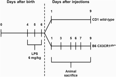
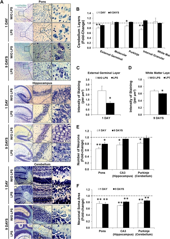
Methods: Systemic inflammation was induced by daily intraperitoneal administration (i.p.) of LPS (6 mg/kg) in newborn mice from postnatal day (PND) 4 to PND6. The effects on neurodevelopment were examined by staining the white matter and neurons with Luxol Fast Blue and Cresyl Violet, respectively. The inflammatory response was assessed by quantifying the expression/activity of matrix metalloproteinases (MMP), toll-like receptor (TLR)-4, high mobility group box (HMGB)-1, and autotaxin (ATX). In addition, B6 CX3CR1(gfp/+) mice combined with cryo-immunofluorescence were used to determine the acute, delayed, and lasting effects on myelination, microglia, and astrocytes.

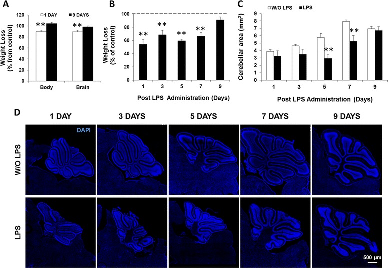
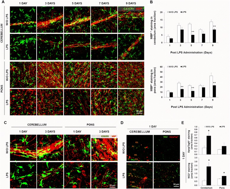
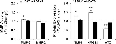
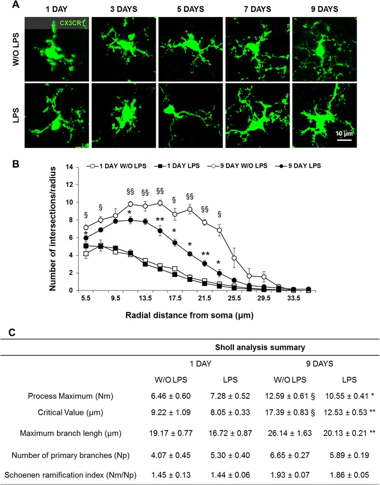
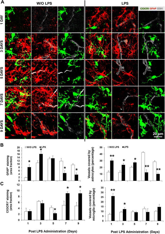
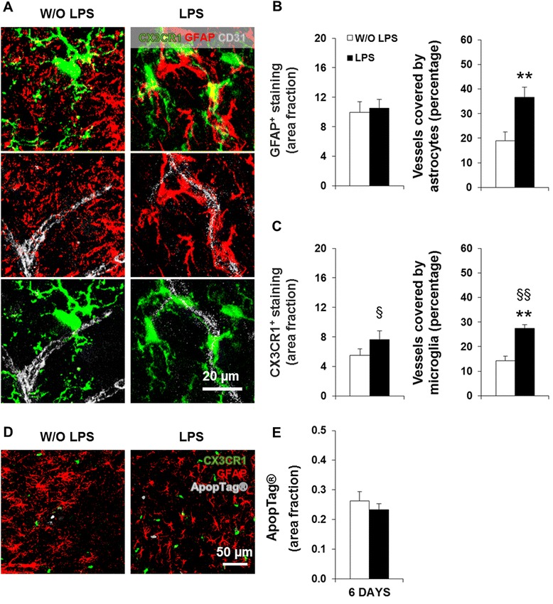
Results: LPS administration led to acute body and brain weight loss as well as overt structural changes in the brain such as cerebellar hypoplasia, neuronal loss/shrinkage, and delayed myelination. The impaired myelination was associated with alterations in the proliferation and differentiation of NG2 progenitor cells early after LPS administration, rather than with excessive phagocytosis by CNS myeloid cells. In addition to disruptions in brain architecture, a robust inflammatory response to LPS was observed. Quantification of inflammatory biomarkers revealed decreased expression of ATX with concurrent increases in HMGB1, TLR-4, and MMP-9 expression levels. Acute astrogliosis (GFAP(+) cells) in the brain parenchyma and at the microvasculature interface together with parenchymal microgliosis (CX3CR1(+) cells) were also observed. These changes preceded the migration/proliferation of CX3CR1(+) cells around the vessels at later time points and the subsequent loss of GFAP(+) astrocytes.

Conclusion: Collectively, our study has uncovered a complex innate inflammatory reaction and associated structural changes in the brains of neonatal mice challenged peripherally with LPS. These findings may explain some of the neurobehavioral abnormalities that develop following neonatal sepsis.


Figures

Figure 1
Figure 1
Schematic representation of the early induction of systemic inflammation. Offspring of both genders were randomly divided into two groups and treated with three intraperitoneal injections of either saline solution or lipopolysaccharide (LPS) at days 4, 5, and 6 after birth to induce systemic inflammation. CD1 wild-type mice were sacrificed at 1 and 9 days after LPS injections, and C57BL/6 (B6) CX3CR1gfp/+ mice were sacrificed at days 1/3/5/6/7/9 following LPS administration.
Figure 2
Figure 2
Early lipopolysaccharide (LPS) administration causes transient body and brain weight loss and decreases the cerebellar area. (A) Body and brain of CD1 wild-type mice were weighed at 1 and 9 days post-LPS administration. (B) Body weight of C57BL/6 (B6) CX3CR1gfp/+ mice was assessed at days 1/3/5/7/9 after LPS injection. (C) Cerebellar area was measured in tiled confocal images of brain sections from B6 CX3CR1gfp/+ mice at days 1/3/5/7/9 post-LPS (representative images with DAPI in (D). Results are mean ± SEM from at least four animals. **P < 0.01 vs. without (W/O) LPS.
Figure 3
Figure 3
Lipopolysaccharide (LPS) administration triggers transient shrinkage of cerebellar layers, acute neuronal loss, and sustained atrophy. Paraffin sections from CD1 wild-type mice at 1 and 9 days post-LPS administration were stained with Luxol Fast Blue (myelin, blue) followed by Cresyl Violet (Nissl bodies, purple). (A) Representative images are shown for each condition in the pons, hippocampus, and cerebellum. (B) The widths of each cerebellar layer [external germinal layer (EGL), molecular layer (ML), Purkinje layer, internal granular layer (IGL), white matter layer (WML)] were measured. (C,D) Intensity of Cresyl Violet staining in the EGL and of Luxol Fast Blue in WML were quantified per square micrometer at 1 and 9 days after LPS administration, respectively. (E,F) The number of neurons per field and the area of neuronal cell body (soma) was quantified throughout the pons, in the CA3 hippocampal region, and in the cerebellar Purkinje layer (PL). All determinations were done using ImageJ software (NIH, USA). Results are mean ± SEM from at least five animals. *P < 0.05 vs. without (W/O) LPS.
Figure 4
Figure 4
Lipopolysaccharide (LPS) administration disrupts myelination in the cerebellum and pons of neonatal mice. Brain cryosections from C57BL/6 CX3CR1gfp/+ mice at days 1/3/5/7/9 post-LPS administration were immunolabeled for myelin (myelin basic protein, MBP, red). Representative confocal images of the cerebellum and pons are displayed in (A), where the overlapping of MBP and microglial marker CX3CR1 (green) is seen in yellow. (B) Area fraction per field of MBP staining was quantified per region using ImageJ software (NIH, USA). (C) To assess microglial phagocytosis, confocal images at days LPS1 and LPS3 were amplified (overlapping of MBP and CX3CR1 is visible in yellow). Sections of animals at LPS1 were immunolabeled for apoptosis (ApopTag, red) and oligodendrocytes precursor cells (NG2 cells, green). (D) Cerebellar layers are identified [external germinal layer (EGL), molecular layer (ML), Purkinje layer, internal granular layer (IGL)]. (E) Area fraction per field of ApopTag and of NG2 positive stainings were determined using ImageJ software (NIH, USA). *P < 0.05 and **P < 0.01 vs. without (W/O) LPS.
Figure 5
Figure 5
Lipopolysaccharide (LPS) challenge promotes differential expression of inflammatory biomarkers in the brain. Whole brain lysates of CD1 wild-type mice at 1 day post-lipopolysaccharide (LPS) administration were used to determine the activities of metalloproteinase(MMP)-9 and MMP-2 by gelatin zymography, alongside with the expression of toll-like receptor (TLR)-4, high-mobility group box 1 (HMGB1) and autotaxin (ATX) by western blot. Results are expressed as fold-change from animals without (W/O) LPS, and are mean ± SEM from at least four animals in each group. *P < 0.05 and **P < 0.01 vs. W/O LPS.
Figure 6
Figure 6
Morphological changes in microglia following lipopolysaccharide (LPS) administration. (A) Representative confocal images of CX3CR1+ cell morphology from brain cryosections of C57BL/6 CX3CR1gfp/+ mice at days 1/3/5/7/9 post-lipopolysaccharide (LPS) administration are shown. (B,C) Using ImageJ software (NIH, USA), we performed Scholl analysis (>40 cells per animal) at days 1 and 9 post-LPS administration. Results are mean ± SEM from at least four animals. *P < 0.05 and **P < 0.01 vs. without (W/O) LPS; § P < 0.05 and §§ P < 0.01 vs. day 1 post-LPS.
Figure 7
Figure 7
Early increased vessel coverage by glia is followed by delayed astrocytic loss and parenchymal microgliosis. Brain cryosections of C57BL/6 CX3CR1gfp/+ mice at days 1/3/5/7/9 post-lipopolysaccharide (LPS) administration were immunolabeled for astrocytes (glial fibrillary acidic protein, GFAP, red) along with vessel marker CD31 (cluster of differentiation 31, gray). Representative confocal images of glia-endothelium interactions in pons are displayed in (A). (B,C) Area fraction per field of GFAP and CX3CR1 (green) positive staining, as well as their respective colocalization with the vessels were determined by ImageJ software (NIH, USA). Results are mean ± SEM from at least four animals. *P < 0.05 and **P < 0.01 vs. without (W/O) LPS.
Figure 8
Figure 8
Astrocytic loss in neonatal inflammation is preceded by proliferation/migration of CX3CR1+ cells in the pons. Brain cryosections of C57BL/6 CX3CR1gfp/+ mice at day 6 post-lipopolysaccharide (LPS) administration were immunolabeled for astrocytes (glial fibrillary acidic protein, GFAP, red) and brain microvascular endothelial cells (cluster of differentiation, CD31, gray). Representative confocal images of each condition in the pons are depicted in (A). (B,C) Area fraction per field of GFAP and CX3CR1 (green) positive staining, along with their respective colocalization with vessels, were determined using ImageJ software (NIH, USA). Representative confocal images of sections immunolabeled for astrocytes (GFAP, red) and for ApopTag (gray) are depicted in (D). Quantification of ApopTag+ staining area fraction, using the abovementioned software, is shown in (E). Results are mean ± SEM from at least four animals. **P < 0.01 vs. without (W/O) LPS; § P < 0.05 and §§ P < 0.01 vs. day 5 post-LPS.

References

    1. Martin GS. Sepsis, severe sepsis and septic shock: changes in incidence, pathogens and outcomes. Expert Rev Anti Infect Ther. 2012;10(6):701–6. - PMC - PubMed
    1. Ogunlesi TA, Ogunfowora OB. Predictors of mortality in neonatal septicemia in an underresourced setting. J Natl Med Assoc. 2010;102(10):915–21. - PubMed
    1. Black RE, Cousens S, Johnson HL, Lawn JE, Rudan I, Bassani DG, et al. Global, regional, and national causes of child mortality in 2008: a systematic analysis. Lancet. 2010;375(9730):1969–87. - PubMed
    1. Zea-Vera A, Ochoa TJ. Challenges in the diagnosis and management of neonatal sepsis. J Trop Pediatr. 2015;61(1):1–13. - PMC - PubMed
    1. Mukherjee R, McQuinn TC, Dugan MA, Saul JP, Spinale FG. Cardiac function and circulating cytokines after endotoxin exposure in neonatal mice. Pediatr Res. 2010;68(5):381–6. - PMC - PubMed

Publication types

MeSH terms

Supplementary concepts