. 2015 Apr 30:15:67.
doi: 10.1186/s12871-015-0050-8.

Comparison of the effects of moderate and severe hypercapnic acidosis on ventilation-induced lung injury

Affiliations

Comparison of the effects of moderate and severe hypercapnic acidosis on ventilation-induced lung injury

Wanchao Yang et al. BMC Anesthesiol. .

Abstract

Background: We have proved that hypercapnic acidosis (a PaCO2 of 80-100 mmHg) protects against ventilator-induced lung injury in rats. However, there remains uncertainty regarding the appropriate target PaCO2 or if greater CO2 "doses" (PaCO2 > 100 mmHg) demonstrate this effect. We wished to determine whether severe acute hypercapnic acidosis can reduce stretch-induced injury, as well as the role of nuclear factor-κB (NF-κB) in the effects of acute hypercapnic acidosis.

Methods: Fifty-four rats were ventilated for 4 hours with a pressure-controlled ventilation mode set at a peak inspiratory pressure (PIP) of 30 cmH2O. A gas mixture of carbon dioxide with oxygen (FiCO2 = 4-5%, FiCO2 = 11-12% or FiCO2 = 16-17%; FiO2 = 0.7; balance N2) was immediately administered to maintain the target PaCO2 in the NC (a PaCO2 of 35-45 mmHg), MHA (a PaCO2 of 80-100 mmHg) and SHA (a PaCO2 of 130-150 mmHg) groups. Nine normal or non-ventilated rats served as controls. The hemodynamics, gas exchange and inflammatory parameters were measured. The role of NF-κB pathway in hypercapnic acidosis-mediated protection from high-pressure stretch injury was then determined.

Results: In the NC group, high-pressure ventilation resulted in a decrease in PaO2/FiO2 from 415.6 (37.1) mmHg to 179.1 (23.5) mmHg (p < 0.001), but improved by MHA (379.9 ± 34.5 mmHg) and SHA (298.6 ± 35.3 mmHg). The lung injury score in the SHA group (7.8 ± 1.6) was lower than the NC group (11.8 ± 2.3, P < 0.05) but was higher than the MHA group (4.4 ± 1.3, P < 0.05). Compared with the NC group, after 4 h of high pressure ventilation, the MHA and SHA groups had decreases in MPO activity of 67% and 33%, respectively, and also declined the levels of TNF-α (58% versus 72%) and MIP-2 (76% versus 60%) in the BALF. Additionally, both hypercapnic acidosis groups reduced stretch-induced NF-κB activation (p < 0.05) and significantly decreased lung ICAM-1 expression (p < 0.05).

Conclusions: Moderate hypercapnic acidosis (PaCO2 maintained at 80-100 mmHg) has a greater protective effect on high-pressure ventilation-induced inflammatory injury. The potential mechanisms may involve alterations in NF-κB activity.


Figures

Figure 1
Figure 1
Effect of hypercapnic acidosis on major markers in BALF in rats’ lungs induced by high-pressure ventilation (HPV) for 4 h. (A) Bronchoalveolar lavage fluid (BALF) total protein levels (microgram/ml) were augmented by high-pressure ventilation but diminished by both hypercapnic groups. (B, C) The total and neutrophil cell count in the lavage group increased following HPV but were attenuated in the hypercapnic groups. (D) Pulmonary edema formation was quantified by measuring the wet and desiccated dry weights of the lung tissue. n = 8 for each group.ap < 0.05 versus the NV group;bp < 0.05 versus the NC group;cp < 0.05 in the SHA group versus the MHA group.
Figure 2
Figure 2
Histologic analysis of lungs. (A and a) NV group; (B and b) NC group; (C and c) MHA group; (D and d) SHA group and (E) lung injury scores in the four groups. NV = normal-pressure ventilation with normocapnia; NC = high-pressure ventilation with normocapnia; MHA = moderate hypercapnic acidosis; SHA = severe hypercapnic acidosis. The A, B, C and D panels represent 100x magnification, and the a, b, c and d panels represent 400x magnification. Severe edema in the alveolar septum and spaces with hyaline membrane formation (red arrow) were seen in the NC group, and rare neutrophil infiltration, moderate interstitial edema and less hyaline membrane formation were observed in both hypercapnic acidosis groups. Horizontal bars represent the median. n = 12 for each group.ap <0.05 versus the NV group; bp <0.05 versus the NC group; cp < 0.001 in the SHA group versus the MHA group.
Figure 3
Figure 3
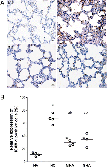
Lung immunohistochemistry for ICAM-1 protein expression in lung tissues. NV = normal-pressure ventilation with normocapnia; NC = high-pressure ventilation with normocapnia; MHA = moderate hypercapnic acidosis; SHA = severe hypercapnic acidosis (A) Immunohistochemical staining of ICAM-1 in the lung. Sections were stained with a brown DAB color-developing agent and counterstained with hematoxylin. The presence of brown granules in the nucleus was defined as a positive cell. All panels represent a 400x magnification. (B) Scatter plot of ICAM-1-positive (+) cells (%) in lung tissue. Semi-quantitative analysis of ICAM-1 indicated that hypercapnic acidosis decreased the expression of ICAM-1. Horizontal bars represent the median. n = 4 for each group. ap <0.05 versus the NV group; bp < 0.05 versus the NC group.
Figure 4
Figure 4
Western blot analysis of the nuclear p65, cytoplasmic IκB-α and ICAM-1 protein in lung tissues. sham = anaesthetized and non-ventilated rats; NC = high-pressure ventilation with normocapnia; MHA = moderate hypercapnic acidosis; SHA = severe hypercapnic acidosis. (A) A representative western blot of lung tissue nuclear p65, cytoplasmic IκBα and ICAM-1 from a sham (unventilated) animal, and animals exposed to HPV under HCA and normocapnic conditions. Both moderate and severe hypercapnic acidosis reduced nuclear p65 expression after 4 h of HPV (p < 0.05). IκB-α fractions were only reduced by HPV and were protected by the use of hypercapnic acidosis (p < 0.05). ICAM-1 showed positive expression, but this was highest in animals treated with only HPV. Densitometric readings of the western blot expressed as NF-κB and IκB-α and ICAM-1/loading control β-actin (panels B, C and D). Horizontal bars represent the median. n = 4 for each group. ap < 0.05 versus the sham group;bp < 0.05 versus the NC group;cp < 0.001 in the SHA group versus the MHA group.
Figure 5
Figure 5
Lung Immunohistochemistry for NF-κB p65 protein expression in lung tissues. NV = normal-pressure ventilation with normocapnia; NC = high-pressure ventilation with normocapnia; MHA = moderate hypercapnic acidosis; SHA = severe hypercapnic acidosis. (A and B) arrows indicate the position of NF-κB p65 expression, which was only observed in the nuclei of airway epithelial cells and alveoli of HPV-treated animals. (C) Scatter plot of NF-κB p65 -positive (+) cells (%) in lung tissue. NF-κB expression also increased after 4 h of HPV compared to the normal ventilation group (p < 0.05), but this was attenuated by both moderate and severe hypercapnia. (D) ELISA analysis of total lung tissue NF-κB indicated that the NF-κB activity also increased after 4 h of HPV compared to the normal ventilation group but was attenuated by both moderate and severe hypercapnia. All panels represent a 400x magnification. Horizontal bars represent the median. n = 4 for each group. ap < 0.05 versus the NV group;bp < 0.001 versus the NC group.

Similar articles

Cited by

References

    1. Dreyfuss D, Basset G, Soler P, Saumon G. Intermittent positive-pressure hyperventilation with high inflation pressures produces pulmonary microvascular injury in rats. Am Rev Respir Dis. 1985;132(4):880–4. - PubMed
    1. Dreyfuss D, Saumon G. Ventilator-induced lung injury: lessons from experimental studies. Am J Respir Crit Care Med. 1998;157(1):294–323. doi: 10.1164/ajrccm.157.1.9604014. - DOI - PubMed
    1. Amato MB, Barbas CS, Medeiros DM, Magaldi RB, Schettino GP, Lorenzi-Filho G, et al. Effect of a protective-ventilation strategy on mortality in the acute respiratory distress syndrome. N Engl J Med. 1998;338(6):347–54. doi: 10.1056/NEJM199802053380602. - DOI - PubMed
    1. Maeda YFY, Uchiyama A, Matsuura N, Mashimo T, Nishimura M. Effects of peak inspiratory flow on development of ventilator-induced lung injury in rabbits. Anesthesiology. 2004;101(3):722–8. doi: 10.1097/00000542-200409000-00021. - DOI - PubMed
    1. Laffey JGTM, Engelberts D, Luo X, Yuan S, Keith Tanswell A, Post M, et al. Therapeutic hypercapnia reduces pulmonary and systemic injury following in vivo lung reperfusion. Am J Respir Crit Care Med. 2000;162(6):2287–94. doi: 10.1164/ajrccm.162.6.2003066. - DOI - PubMed

Publication types

MeSH terms