

Treatment of osteonecrosis of the femoral head with focal anatomic-resurfacing implantation (HemiCAP): preliminary results of an alternative option
- PMID: 25924980
- PMCID: PMC4423414
- DOI: 10.1186/s13018-015-0199-3
Treatment of osteonecrosis of the femoral head with focal anatomic-resurfacing implantation (HemiCAP): preliminary results of an alternative option
Abstract
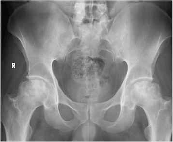
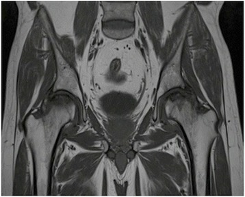
Background: The optimal treatment of osteonecrosis of the femoral head has not been established yet. The aim of this study was to report preliminary clinical results of focal anatomic-resurfacing implantation for the treatment of osteonecrosis of the femoral head.
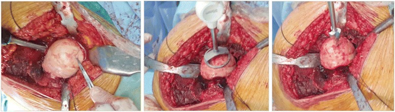
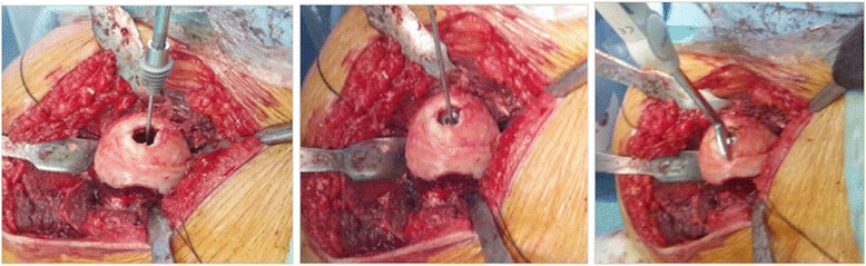
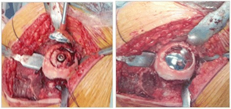
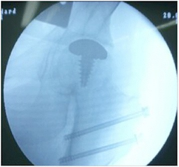
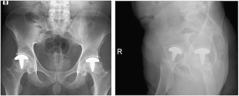
Methods: Five patients (four male, one female) with seven surgical procedures, ages between 37 and 52 with an average age of 45.2 (+/- 7.2), diagnosed as femoral head avascular necrosis and who were unresponsive to conservative management or had failed previous surgical treatments were treated with a focal anatomic femoral head resurfacing between the years 2011-2012 and were retrospectively reviewed. Five patients with at least two years of follow-up, one left hip, two right hips, and two patients with bilateral hip surgery were included in this review. After safe surgical dislocation of the hip, full exposure of the femoral head was established. A focal-resurfacing implant matching patient anatomy and femoral head curvature was performed accordingly. Neither intraoperative or postoperative complications nor revision ensued. Visual analogue scores and Harris Hip Scores were recorded both preoperatively and at postoperative 2 years for all seven surgeries.
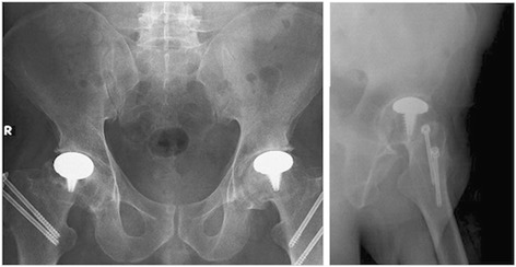
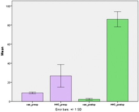
Results: The mean follow-up period was 26.6 +/- 3.8 months, with a range between 24-33 months. The mean visual analogue scores were 8.9 +/- 0.9 preoperatively and 2.3 +/- 1.0 postoperatively at year two (p = 0.017). Harris Hip Scores at postoperative follow-up were found to improve significantly from good to excellent scores (86.0 +/- 7.9), compared with preoperative poor scores (26.7 +/- 11.8) (p = 0.018). The clinical improvements in visual analogue scores (VAS) and Harris Hip Scores were also found to correlate with each other (p < 0.05).
Conclusions: In the present study, the alternative technique of focal anatomic hip resurfacing with HemiCAP® yielded preliminary successful results for the treatment of osteonecrosis of the femoral head. To the best of our knowledge, this is the first case series in the literature, reporting functional clinical results with the use of a focal anatomic-resurfacing implant for the treatment of focal femoral head osteonecrosis.
Figures
References
-
- Mont MA, Hungerfort DS. Non-traumatic avascular necrosis of the femoral head. J Bone Joint Surg Am. 1995;77:459–74. - PubMed
-
- Pivec IK, Kapadia BH, Banerjee S, Mont MA. Osteonecrosis of the femoral head. Bone Joint J. 2013;95-B(Suppl A):46–50002E. - PubMed
-
- Lee JS, Lee JS, Rob HL, Kim CH, Js J, Suh KT. Alteration in the differentiation ability of mesenchymal stem cells in patients with nontraumatic osteonecrosis of the femoral head: comparative analysis according to the risk factor. J Orthop Res. 2006;24(4):604–9. doi: 10.1002/jor.20078. - DOI - PubMed
MeSH terms
LinkOut - more resources
Full Text Sources
Other Literature Sources
