. 2015 Jun 15;308(12):F1452-62.
doi: 10.1152/ajprenal.00066.2015. Epub 2015 Apr 29.

Muc1 is protective during kidney ischemia-reperfusion injury

Affiliations

Muc1 is protective during kidney ischemia-reperfusion injury

Núria M Pastor-Soler et al. Am J Physiol Renal Physiol. .

Abstract

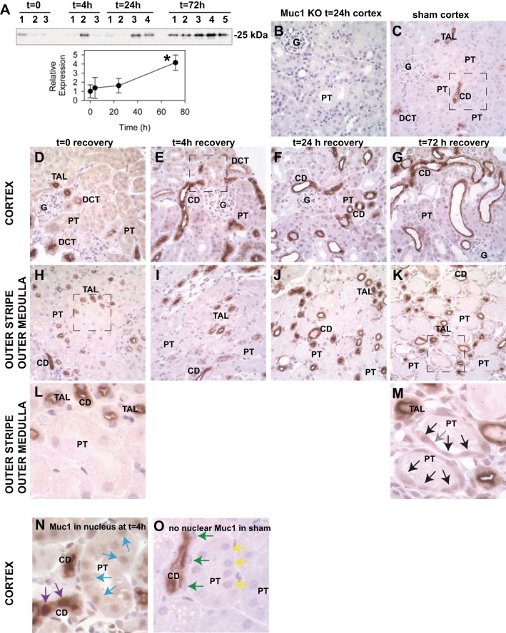
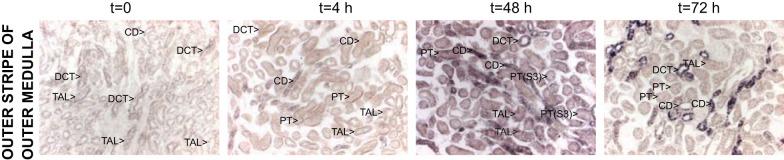
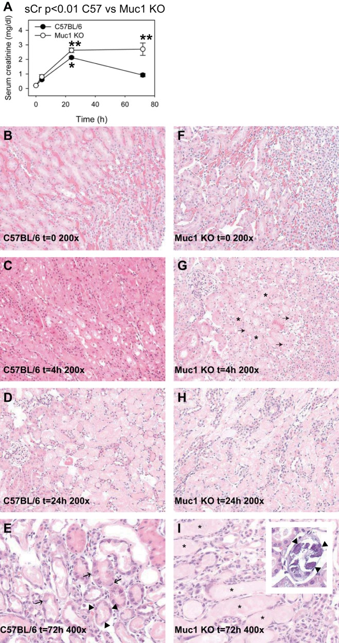
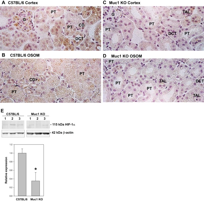
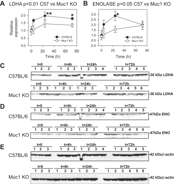
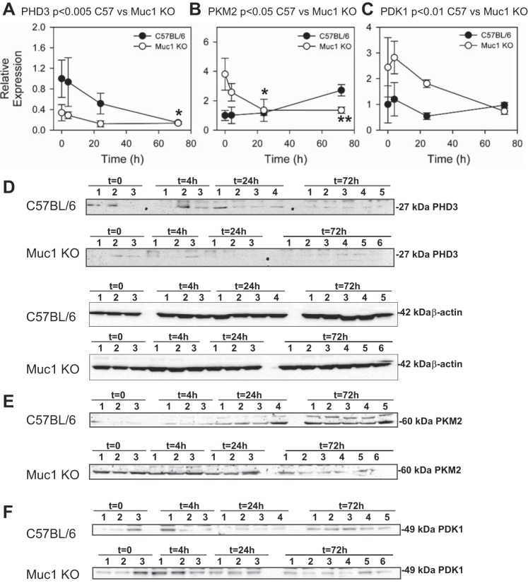
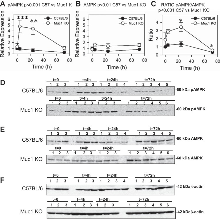
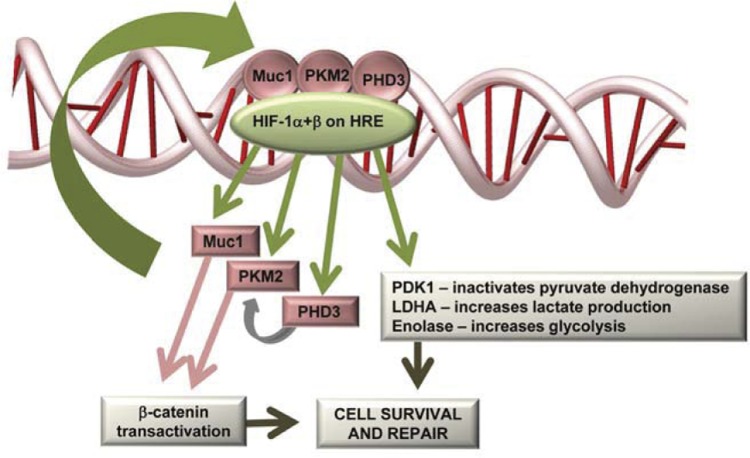
Ischemia-reperfusion injury (IRI) due to hypotension is a common cause of human acute kidney injury (AKI). Hypoxia-inducible transcription factors (HIFs) orchestrate a protective response in renal endothelial and epithelial cells in AKI models. As human mucin 1 (MUC1) is induced by hypoxia and enhances HIF-1 activity in cultured epithelial cells, we asked whether mouse mucin 1 (Muc1) regulates HIF-1 activity in kidney tissue during IRI. Whereas Muc1 was localized on the apical surface of the thick ascending limb, distal convoluted tubule, and collecting duct in the kidneys of sham-treated mice, Muc1 appeared in the cytoplasm and nucleus of all tubular epithelia during IRI. Muc1 was induced during IRI, and Muc1 transcripts and protein were also present in recovering proximal tubule cells. Kidney damage was worse and recovery was blocked during IRI in Muc1 knockout mice compared with congenic control mice. Muc1 knockout mice had reduced levels of HIF-1α, reduced or aberrant induction of HIF-1 target genes involved in the shift of glucose metabolism to glycolysis, and prolonged activation of AMP-activated protein kinase, indicating metabolic stress. Muc1 clearly plays a significant role in enhancing the HIF protective pathway during ischemic insult and recovery in kidney epithelia, providing a new target for developing therapies to treat AKI. Moreover, our data support a role specifically for HIF-1 in epithelial protection of the kidney during IRI as Muc1 is present only in tubule epithelial cells.

Keywords: AMP-activated protein kinase; Muc1; acute kidney injury; hypoxia-inducible factor-1 (HIF-1); ischemia.


Figures

Fig. 1.
Fig. 1.
Mucin (Muc)1 is induced during kidney ischemia-reperfusion injury (IRI) and appears in the nucleus. Kidneys of control C57BL/6 mice were subjected to 19 min of ischemia and recovery for 0–72 h (n = 3–5 mice at each time point). A: immunoblot analysis of control mouse kidney homogenates (60 μg protein/lane) with anti-Muc1 cytoplasmic tail antibodies (small subunit Mr: 25 kDa) and then anti-β-actin antibodies as a loading control (Fig. 6D) revealed a significant 4.2-fold increase of Muc1 by 72 h using one-way ANOVA (means ± SE, *P < 0.05). B–O: slices of paraformaldehyde (PFA)-fixed tissue were subjected to immunohistochemistry with the same anti-Muc1 antibody. Images for B–K are ×400 magnification. B: no Muc1 signal was observed in kidneys of Muc1 knockout (KO) mice, as expected (negative control). C: renal Muc1 staining in sham-treated C57BL/6 mice was predominantly in the thick ascending limb (TAL), distal convoluted tubule (DCT), and collecting duct (CD) (box enlarged in O). After 19 min of ischemia [time (t) = 0], Muc1 staining was also apparent in the cytoplasm of the TAL, DCT, CD, and proximal tubules (PTs) in the cortex (D) and outer stripe of the outer medulla (H and L). At 4, 24, and 72 h of recovery, Muc1 staining was also apparent in the nucleus (E–G and I–K). Boxes in H and K are enlarged in L and M, respectively, to emphasize Muc1 staining in the cytoplasm and nucleus of recovering PT (black arrows) and on the cell surface (gray arrow). Nuclear staining was clearly evident in the CD (purple arrows) and PT (blue arrows) at 4 h of recovery and absent in the CD (green arrows) and PT (yellow arrows) in sham-treated mice (compare N and O). G, glomerulus.
Fig. 2.
Fig. 2.
Muc1 transcripts are induced during kidney IRI. Kidneys of control C57BL/6 mice were subjected to IRI as described in Fig. 1. PFA-fixed kidneys were subjected to in situ hybridization using a Muc1-specific probe. Transcripts for Muc1 identified with in situ hybridization were evident at t = 0 but notably increased by 4 h of recovery in the TAL, DCT, and CD [outer stripe of the outer medulla (OSOM) is shown]. Transcripts were maximally increased at 48 h of recovery in all tubules, including the PT [see label for PT(S3) as shown in the third panel 48 h].
Fig. 3.
Fig. 3.
Muc1 protects kidney function and morphology during kidney IRI. Kidneys of Muc1 KO mice and congenic control C57BL/6 mice were subjected to IRI for 19 min and recovery for 0–72 h. A: blood was collected at the time of death and assayed for serum creatinine (sCr). Profiles for sCr between Muc1 KO mice (open circles) and congenic wild-type mice (closed circles) were significantly different by two-way ANOVA (n = 3–6 mice at each time point, P < 0.01). Values at 72 h (means ± SE) were different between Muc1 KO and wild-type mice (P < 0.001). Values at 24 h in control mouse kidneys were significantly higher than at t = 0 (*P < 0.01). Values for Muc1 KO mouse kidneys at both 24 and 72 h were significantly higher than at t = 0 (**P < 0.001). B–I: one kidney from each mouse was fixed and stained with hematoxylin and eosin and scored for tubule damage as described in the text. B–H: representative images (×200) are shown for control (B–D) and Muc1 KO (F–H) kidneys at 0, 4, and 24 h of recovery, respectively. G: karyorrhexis and karyopyknosis (arrows) and luminal casts (*) were especially evident in Muc1 KO kidneys at t = 4 h. E and I: images (×400) from 72 h of recovery are shown for control C57BL/6 (E) and Muc1 KO (I) mice. E: mitotic figures (arrows) and flattened cells in recovering tubules (arrow heads) were observed in control mouse kidneys at 72 h. I and inset: luminal casts (*) and calcium phosphate precipitates (arrowheads) were observed in Muc1 KO kidneys at 72 h.
Fig. 4.
Fig. 4.
Muc1 stabilizes hypoxia-inducible factor (HIF)-1α levels during IRI. Kidneys of Muc1 KO mice and congenic control C57BL/6 mice were subjected to IRI for 19 min and recovery for 4 h. A–D: one kidney from each mouse was fixed and stained by immunohistochemistry with anti-HIF-1α antibody. Levels of HIF-1α were notably reduced in the Muc1 KO mouse cortex and OSOM (C and D) compared with the control mouse kidney (A and B). Examples of the glomerulus (G), PT, DCT, TAL, and CD are shown. E: immunoblot analysis of mouse kidney homogenates (60 μg protein/lane) with anti-HIF-1α antibodies revealed a significant decrease in the absence of Muc1 using Student's t-test (means ± SE, n = 3, *P < 0.05). Immunoblot analysis of β-actin was used as a loading control.
Fig. 5.
Fig. 5.
Muc1 transactivates gene targets of HIF-1: lactate dehydrogenase A (LDHA) and enolase (ENO). Kidneys of Muc1 KO mice and congenic control C57BL/6 mice were subjected to IRI for 19 min and recovery for 0–72 h. Products of HIF-1 gene targets were measured by immunoblot analysis of 60 μg kidney homogenates for LDHA (A and C) and ENO (B and D) and then β-actin as a loading control (E). Bands on immunoblots were quantified and presented as means ± SE relative to that of control mice at t = 0 (set as 1). Profiles of LDHA and ENO for Muc1 KO and control mouse kidneys were significantly different by two-way ANOVA (P < 0.01 and P < 0.05, respectively, as indicated). Levels of LDHA were significantly increased in control mice at 24 h (**P < 0.01) and 72 h (*P < 0.05) of recovery compared with 0 h. Levels of LDHA at 4, 24, and 72 h of recovery were significantly different between Muc1 KO and control mice (P < 0.05). Levels of ENO were significantly increased in control mice at 24 h (*P < 0.05). Levels of ENO at 24 h were significantly different between Muc1 KO and control mice (P < 0.05).
Fig. 6.
Fig. 6.
Response of gene targets of HIF-1 are Muc1 dependent: prolyl hydroxylase domain 3 (PHD3), pyruvate kinase M2 (PKM2), and pyruvate dehydrogenase kinase 1 (PDK1). Kidneys of Muc1 KO mice and congenic control C57BL/6 mice were subjected to IRI for 19 min and recovery for 0–72 h. Products of three HIF-1 gene targets were measured by immunoblot analysis 60 μg kidney homogenates for PHD3 (A and D), PKM2 (B and E), and PDK1 (C and F). Immunoblot analysis of β-actin was used as a loading control (included in D for PHD3 and shown in Fig. 5E for PKM2 and PDK1). Bands on immunoblots were quantified and presented as means ± SEM relative to that of control mice at t = 0 (set as 1). Profiles of PHD3 (P < 0.005), PKM2 (P < 0.05), and PDK1(P < 0.01) for Muc1 KO and control mouse kidneys were significantly different by two-way ANOVA. Levels of PHD3 were significantly different between control and Muc1 KO mouse kidneys at t = 0 and 4 h of recovery (P < 0.05), and levels of PHD3 in control mouse kidneys at 72 h were significantly less than at t = 0 (*P < 0.05). Levels of PKM2 were significantly different at t = 0 between control and Muc1 KO mouse kidneys (P < 0.01). Levels of PKM2 were significantly reduced in Muc1 KO mouse kidneys after 24 h (*P < 0.05) and 72 h (**P < 0.01) of recovery compared with levels at t = 0.
Fig. 7.
Fig. 7.
Muc1 prevents metabolic stress during IRI. Kidneys of Muc1 KO mice and congenic control C57BL/6 mice were subjected to IRI for 19 min and recovery for 0–72 h. Metabolic stress was assessed by immunoblot analysis of 60 μg kidney homogenates for activated phosphorylated AMP-activated protein kinase (pAMPK; A and D), AMPK (B and E), and β-actin (F) as a loading control. Bands on immunoblots were quantified and presented as means ± SE relative to that of control mice at t = 0 (set as 1). Data are also presented as the ratio of pAMPK to AMPK (C). Profiles of pAMPK (P < 0.001), AMPK (P < 0.01), and the ratio of pAMPK to AMPK (P < 0.001) for Muc1 KO and control mouse kidneys were significantly different by two-way ANOVA. Levels of pAMPK in Muc1 KO mice were significantly increased at 4 h (***P < 0.001) and 24 h (**P < 0.005) of recovery compared with levels at t = 0 and significantly higher at 4 and 24 h of recovery than levels for control kidney levels (P < 0.001). Levels of AMPK in Muc1 KO kidneys were significantly higher at 24 and 72 h (P < 0.05) than levels in control kidneys. The ratio of pAMPK to AMPK in Muc1 KO mice was significantly different at 24 and 72 h of recovery compared with levels at t = 0 (*P < 0.05), and the ratio was significantly higher than in control mouse kidneys at 4 and 24 h of recovery (P < 0.005).
Fig. 8.
Fig. 8.
Muc1 transactivates the HIF-1 protective pathway during IRI in the kidney. HIF-1, composed of an oxygen-regulated α-subunit and a constitutively expressed β-subunit, binds to hypoxia-responsive elements (HREs) and promotes transcription of 1) genes to shift glucose metabolism; 2) PHD3, which hydroxylates PKM2; 3) PKM2, which transactivates both HIF-1 and β-catenin; and 4) Muc1, which binds, stabilizes, and transactivates both β-catenin and HIF-1 protective pathways. See text for further discussion.

Similar articles

Cited by

References

    1. Adkison D, Hollwarth ME, Benoit JN, Parks DA, McCord JM, Granger DN. Role of free radicals in ischemia-reperfusion injury to the liver. Acta Physiol Scand Suppl 548: 101–107, 1986. - PubMed
    1. Aubert S, Fauquette V, Hemon B, Lepoivre R, Briez N, Bernard D, Van Seuningen I, Leroy X, Perrais M. MUC1, a new hypoxia inducible factor target gene, is an actor in clear renal cell carcinoma tumor progression. Cancer Res 69: 5707–5715, 2009. - PubMed
    1. Bernhardt WM, Campean V, Kany S, Jurgensen JS, Weidemann A, Warnecke C, Arend M, Klaus S, Gunzler V, Amann K, Willam C, Wiesener MS, Eckardt KU. Preconditional activation of hypoxia-inducible factors ameliorates ischemic acute renal failure. J Am Soc Nephrol 17: 1970–1978, 2006. - PubMed
    1. Bleyer AJ, Kmoch S, Antignac C, Robins V, Kidd K, Kelsoe JR, Hladik G, Klemmer P, Knohl SJ, Scheinman SJ, Vo N, Santi A, Harris A, Canaday O, Weller N, Hulick PJ, Vogel K, Rahbari-Oskoui FF, Tuazon J, Deltas C, Somers D, Megarbane A, Kimmel PL, Sperati CJ, Orr-Urtreger A, Ben-Shachar S, Waugh DA, McGinn S, Bleyer AJ Jr, Hodanova K, Vylet'al P, Zivna M, Hart TC, Hart PS. Variable clinical presentation of an MUC1 mutation causing medullary cystic kidney disease type 1. Clin J Am Soc Nephrol 9: 527–535, 2014. - PMC - PubMed
    1. Braga VM, Pemberton LF, Duhig T, Gendler SJ. Spatial and temporal expression of an epithelial mucin, Muc-1, during mouse development. Development 115: 427–437, 1992. - PubMed

Publication types

LinkOut - more resources