. 2015 Apr 29;6(1):85.
doi: 10.1186/s13287-015-0070-9.

Activation and regulation of the granulation tissue derived cells with stemness-related properties

Affiliations

Activation and regulation of the granulation tissue derived cells with stemness-related properties

Zelin Chen et al. Stem Cell Res Ther. .

Abstract

Introduction: Skin as the largest and easily accessible organ of the body represents an abundant source of adult stem cells. Among them, dermal stem cells hold great promise in tissue repair and the skin granulation tissue has been recently proposed as a promising source of dermal stem cells, but their biological characteristics have not been well investigated.

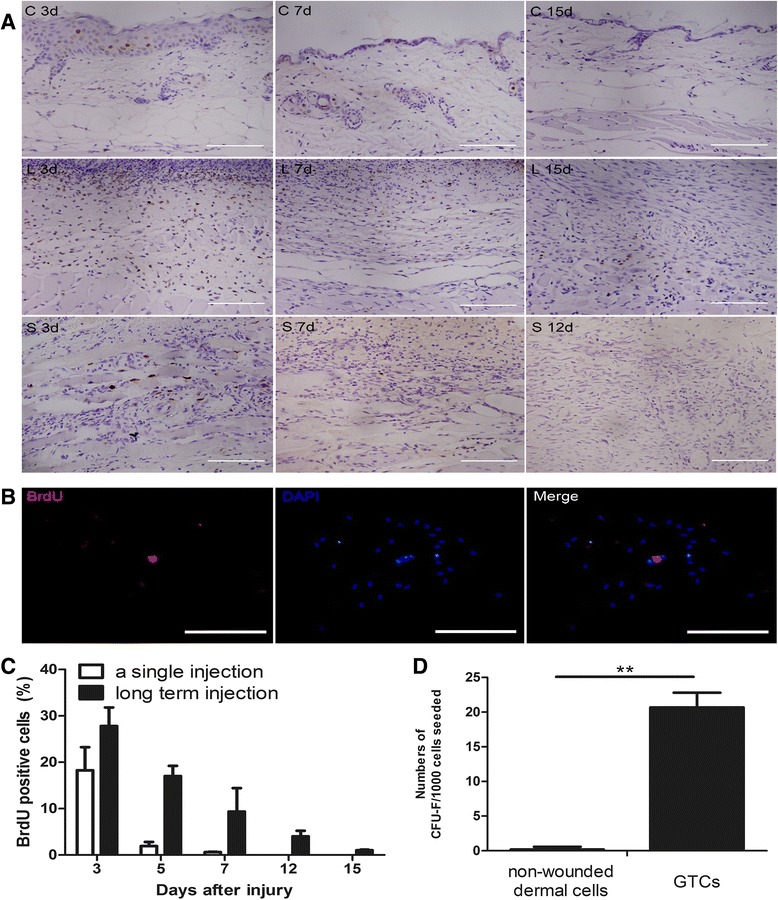
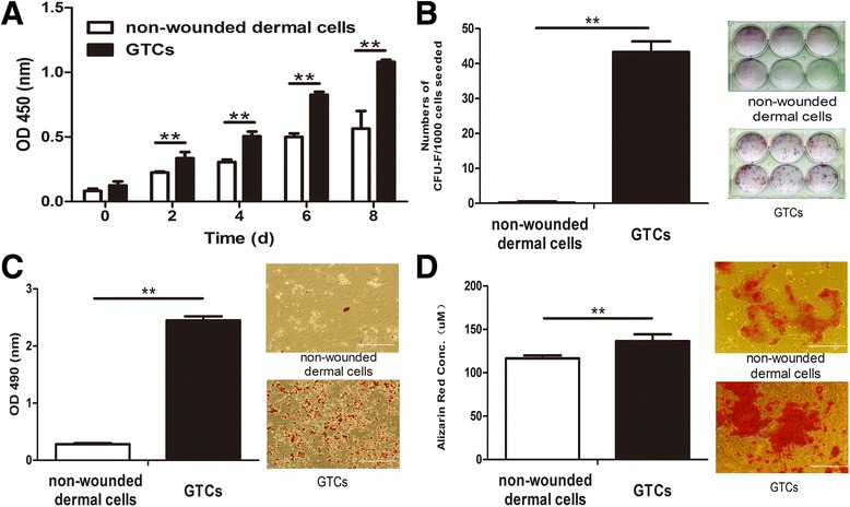
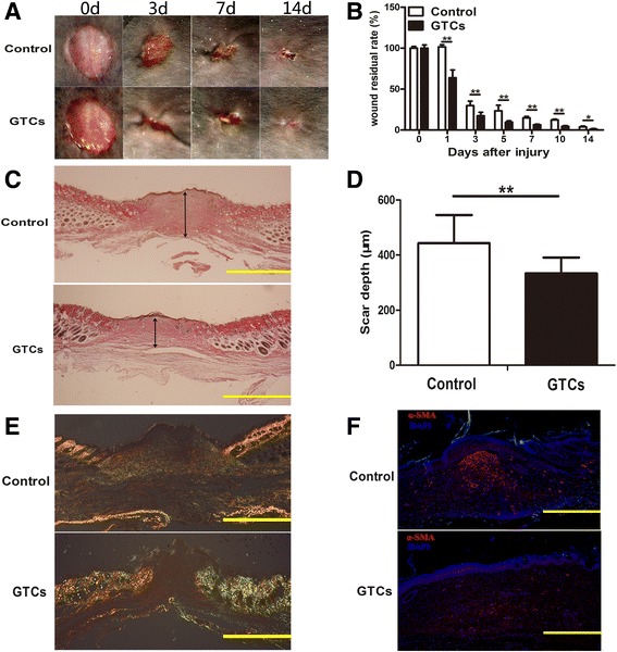
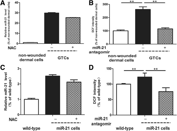
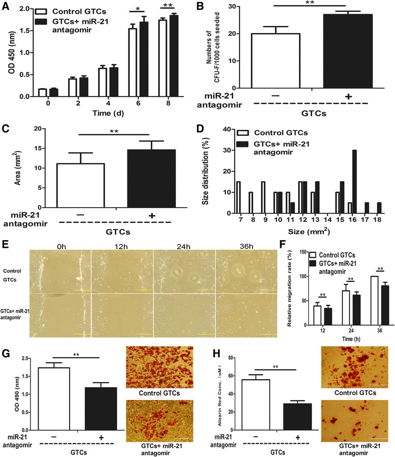
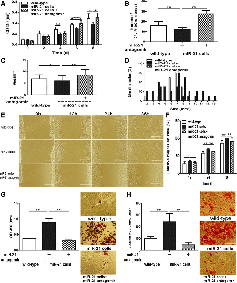
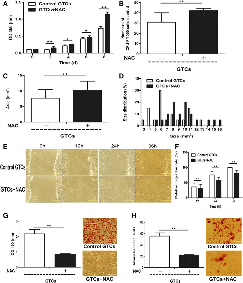
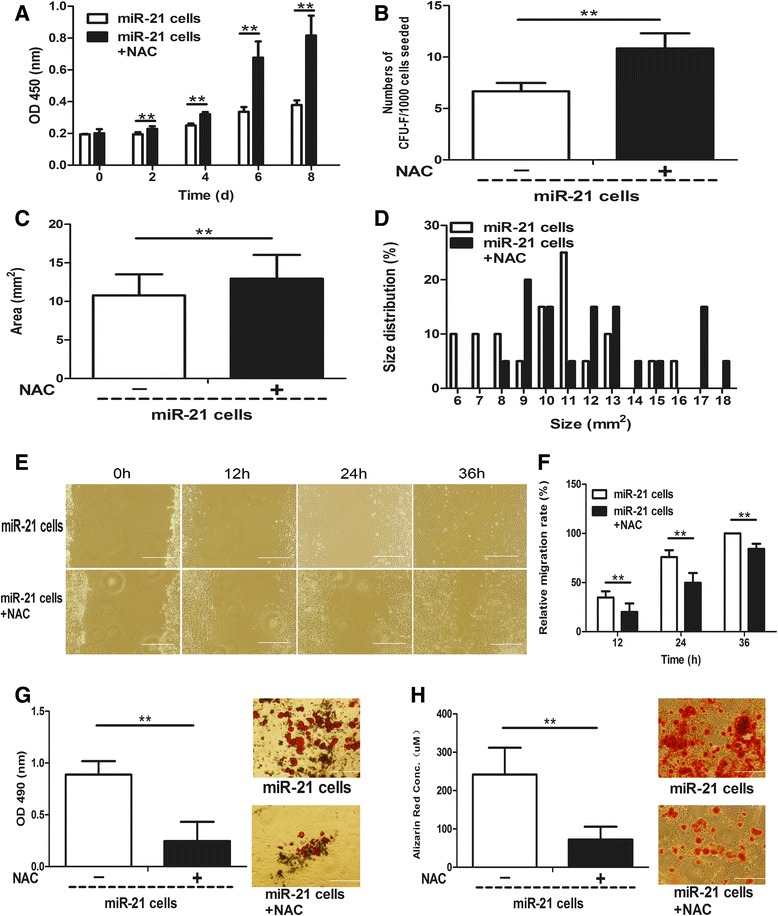
Methods: The 5-bromo-2'-deoxyuridine (BrdU) lineage tracing approach was employed to chase dermal stem cells in vivo. Granulation tissue derived cells (GTCs) were isolated and their in vitro proliferation, self-renewing, migration, and multi-differentiation capabilities were assessed. Combined radiation and skin wound model was used to investigate the therapeutic effects of GTCs. MicroRNA-21 (miR-21) antagomir was used to antagonize miR-21 expression. Reactive oxygen species (ROS) were scavenged by N-acetyl cysteine (NAC).

Results: The quiescent dermal stem/progenitor cells were activated to proliferate upon injury and enriched in granulation tissues. GTCs exhibited enhanced proliferation, colony formation and multi-differentiation capacities. Topical transplantation of GTCs into the combined radiation and skin wound mice accelerated wound healing and reduced tissue fibrosis. Blockade of the miR-21 expression in GTCs inhibited cell migration and differentiation, but promoted cell proliferation and self-renewing at least partially via a ROS dependent pathway.

Conclusions: The granulation tissue may represent an alternative adult stem cell source in tissue replacement therapy and miR-21 mediated ROS generation negatively regulates the stemness-related properties of granulation tissue derived cells.


Figures

Figure 1
Figure 1
Activation of dermal stem/progenitor cell proliferation after wounding. (A) At 3, 5, 7, 12, and 15 days after wounding, 5-bromo-2′-deoxyuridine (BrdU) labels in the normal skin tissues or wounded tissues were detected by immunohistochemistry. The percentage of BrdU-positive cells was also calculated, presented as mean ± standard deviation (C). C, control group (n = 3 per time point); d, days after wounding; L, long-term BrdU labeling group (n = 3 per time point); S, a single BrdU labeling group (n = 3 per time point). Scale bar = 100 μm. (B) BrdU (100 mg/kg) was injected intraperitoneally for three consecutive days (once per 12 hours) in nonwounded or wounded mice. Nonwounded dermal cells or granulation tissue-derived cells (GTCs) at 7 days post wounding were isolated and adherent cells were harvested for colony-forming assay. BrdU labels in the colonies were detected by immunofluorescence. Scale bar = 500 μm. (D) Quantification of the colonies. CFU, colony-forming unit. **P < 0.01.
Figure 2
Figure 2
Stemness-related properties of granulation tissue-derived cells. Passage 2 granulation tissue-derived cells (GTCs) or nonwounded dermal cells were used for experiments. (A) The proliferation ability of GTCs and nonwounded dermal cells was detected every 2 days after seeding. (B) Colony-forming assay of GTCs and nonwounded dermal cells. Results expressed as mean ± standard deviation (n = 6 wells per group). (C) Adipogenic differentiation was induced in mouse mesenchymal stem cell adipogenic differentiation medium. After 22 days, cells were fixed and stained by Oil-Red O. The Oil-Red O staining was also quantified. (D) Osteogenic differentiation was performed in mouse mesenchymal stem cell osteogenic differentiation medium. After 21 days, cells were fixed and stained by Alizarin Red. The Alizarin Red staining was also quantified. d, days; OD, optical density. **P <0.01. Scale bar = 500 μm.
Figure 3
Figure 3
Therapeutic effects of granulation tissue-derived cells on combined radiation and skin wound injury. Granulation tissue-derived cells (GTCs), 1 × 106 per mouse in 0.2 ml phosphate-buffered saline (PBS), were injected around the skin wound margins post 6 Gy total body radiation combined skin wound injury. PBS (0.2 ml per mouse) served as control. (A) Representative wounds injected with PBS (control) or GTCs. (B) Wound residual rates of the GTC-treated group and the control group are presented as mean ± standard deviation (n = 6 mice per group). (C) The fibrotic tissue depths were quantified in serial sections in the center of day 15 wounds treated with PBS or GTCs. (D) Results of the fibrotic tissue depths presented as mean ± standard deviation (n = 6). (E) Representative photomicrographs of Sirius Red-stained sections from day 15 wounds injected with PBS or GTCs. (F) Immunostaining for the classical myofibroblast-specific marker alpha smooth muscle actin (α-SMA) of wound sections derived from day 15 wounds injected with PBS or GTCs. Scale bars = 500 μm. d, days; DAPI, 4′,6-diamidino-2-phenylindole. **P <0.01, *P <0.05.
Figure 4
Figure 4
miR-21 regulates the reactive oxygen species level of dermal-derived cells. (A) Relative microRNA (miR)-21 levels of primary nonwounded dermal cells, granulation tissue-derived cells (GTCs), and GTCs pretreated with N-acetylcysteine (NAC; 5 mM) for 48 hours were tested by quantitative real-time PCR. (B) Relative reactive oxygen species (ROS) levels of primary nonwounded dermal cells, GTCs, and GTCs pretreated with miR-21 antagomir (50 nM) for 48 hours were detected by the ROS-sensitive dye 2′,7′-dichlorofluorescin diacetate (DCFH-DA; 10 mM). (C) Relative miR-21 levels of primary wild-type neonatal dermal cells, miR-21 knock-in neonatal dermal cells, and miR-21 knock-in neonatal dermal cells pretreated with NAC (5 mM) for 48 hours were detected by quantitative real-time PCR. (D) Relative ROS levels of primary wild-type neonatal dermal cells, miR-21 knock-in neonatal dermal cells, and miR-21 knock-in neonatal dermal cells pretreated with miR-21 antagomir (50 nM) for 48 hours were measured by DCFH-DA (10 mM). All results presented as mean ± standard deviation. d, days; DCF, 2′,7′-dichlorofluorescin; h, hours; miR-21 cells, miR-21 knock-in neonatal dermal cells; wild-type, wild-type neonatal dermal cells. **P < 0.01.
Figure 5
Figure 5
miR-21 negatively regulates stemness-related properties of granulation tissue-derived cells. Passage 2 granulation tissue-derived cells (GTCs) were pretreated with microRNA (miR)-21 antagomir (50 nM) or control antagomir (50 nM) for 48 hours. (A) The proliferation ability was detected by CCK-8 every 2 days after seeding. (B) Colony-forming assay of GTCs pretreated with miR-21 antagomir and control antagomir. Colonies presented as mean ± standard deviation (n = 6 wells per group). Twenty colonies were randomly chosen in each group for analyzing (C) average colony sizes and (D) size distribution. (E) Representative migration photographs of GTCs pretreated with miR-21 antagomir and control antagomir at indicated time points. (F) Relative migration rate presented as mean ± standard deviation (n = 6 per time point for each group). (G) Adipogenic and (H) osteogenic differentiation of GTCs pretreated with miR-21 antagomir or control antagomir were measured by staining with Oil-Red O and Alizarin Red and quantifying them respectively. CFU, colony-forming units; d, days; h, hours; OD, optical density. *P < 0.05, **P < 0.01. Scale bar = 500 μm.
Figure 6
Figure 6
miR-21 negatively regulates stemness-related properties of miR-21 knock-in neonatal dermal cells. Passage 2 microRNA (miR)-21 knock-in neonatal dermal cells were pretreated with miR-21 antagomir or control antagomir (50 nM) for 48 hours, and wild-type neonatal dermal cells were treated with control antagomir (50 nM) for 48 hours. (A) The proliferation ability of the three groups was detected every 2 days after seeding. Colony formation assay including (B) colony numbers, (C) colony sizes, and (D) colony size distribution of the three groups was also measured. (E) Representative photographs of migration of the three groups at indicated time points. (F) Relative migration rate of the three groups presented as mean ± standard deviation (n = 6 per time point for each group). (G) Adipogenic and (H) osteogenic differentiation of the three groups were measured by staining with Oil-Red O and Alizarin Red and quantifying them respectively. CFU, colony-forming units; d, days; h, hours; miR-21 cells, miR-21 knock-in neonatal dermal cells; wild-type, wild-type neonatal dermal cells; OD, optical density. *P < 0.05, **P < 0.01. Scale bar = 500 μm.
Figure 7
Figure 7
miR-21 negatively regulates stemness-related properties of granulation tissue-derived cells via a reactive oxygen species-dependent pathway. N-acetyl cysteine (NAC; 5 mM) was added into culture medium and adipogenic and osteogenic induced medium of granulation tissue-derived cells (GTCs) to scavenge the intracellular reactive oxygen species (ROS). (A) Proliferation of GTCs with and without NAC was measured every 2 days after seeding. Colony formation assay including (B) colony numbers, (C) average colony sizes and (D) colony size distribution of GTCs with and without NAC was present. (E) Representative migration photographs of GTCs with and without NAC at indicated time points. (F) The migration rate of the two groups presented as mean ± standard deviation (n = 6 per time point for each group). (G) The quantification and representative photographs of the Oil-Red O staining of the adipogenic differentiation of GTCs with and without NAC. (H) Quantification and representative photographs of the Alizarin Red staining of the osteogenic differentiation of GTCs and GTCs with NAC. CFU, colony-forming units; d, days; h, hours, OD, optical density. *P <0.05; **P <0.01. Scale bar = 500 μm.
Figure 8
Figure 8
miR-21 negatively regulates stemness-related properties of miR-21 knock-in neonatal dermal cells via a reactive oxygen species dependent pathway. N-acetyl cysteine (NAC; 5 mM) was added into culture medium and adipogenic and osteogenic induced medium of miR-21 knock-in neonatal dermal cells to scavenge intracellular reactive oxygen species (ROS), and stemness related-properties of miR-21 knock-in neonatal dermal cells with and without NAC were detected. (A) Proliferation was measured every 2 days after seeding. Colony formation assay including (B) colony numbers, (C) average colony sizes, and (D) colony size distribution was present. (E) Representative migration photographs at indicated time points. (F) Relative migration rate presented as mean ± standard deviation (n = 6 per time point for each group). (G) Quantification and representative photographs of the Oil-Red O staining of the adipogenic differentiation. (H) Quantification and representative photographs of the Alizarin Red staining of the osteogenic differentiation. CFU, colony-forming units; d, days; h, hours; miR-21 cells, miR-21 knock in neonatal dermal cells; OD, optical density. **P <0.01. Scale bar = 500 μm.

Similar articles

Cited by

References

    1. Watt FM. Epidermal stem cells: markers, patterning and the control of stem cell fate. Philos Trans R Soc Lond B Biol Sci. 1998;353:831–7. doi: 10.1098/rstb.1998.0247. - DOI - PMC - PubMed
    1. Hughes S. Epidermal stem cells. J Pathol. 2002;197:479–91. doi: 10.1002/path.1159. - DOI - PubMed
    1. Ghazizadeh S, Taichman LB. Multiple classes of stem cells in cutaneous epithelium: a lineage analysis of adult mouse skin. EMBO J. 2001;20:1215–22. doi: 10.1093/emboj/20.6.1215. - DOI - PMC - PubMed
    1. Uzarska M, Porowinska D, Bajek A, Drewa T. Epidermal stem cells – biology and potential applications in regenerative medicine. Postepy Biochem. 2013;59:219–27. - PubMed
    1. Myung P, Andl T, Ito M. Defining the hair follicle stem cell (Part II) J Cutan Pathol. 2009;36:1134–7. doi: 10.1111/j.1600-0560.2009.01412.x. - DOI - PubMed

Publication types

MeSH terms