. 2015 Jul;42(1):1651-9.
doi: 10.1111/ejn.12935. Epub 2015 May 21.

Automatic representation of a visual stimulus relative to a background in the right precuneus

Affiliations

Automatic representation of a visual stimulus relative to a background in the right precuneus

Motoaki Uchimura et al. Eur J Neurosci. 2015 Jul.

Abstract

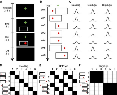
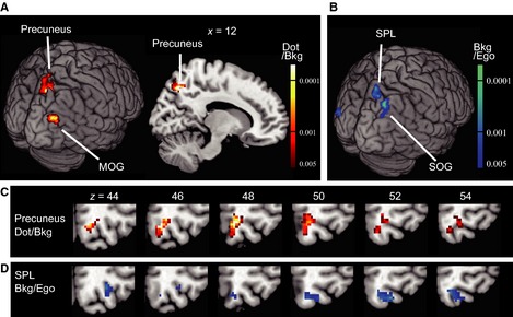
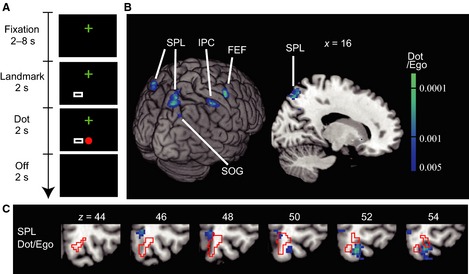
Our brains represent the position of a visual stimulus egocentrically, in either retinal or craniotopic coordinates. In addition, recent behavioral studies have shown that the stimulus position is automatically represented allocentrically relative to a large frame in the background. Here, we investigated neural correlates of the 'background coordinate' using an fMRI adaptation technique. A red dot was presented at different locations on a screen, in combination with a rectangular frame that was also presented at different locations, while the participants looked at a fixation cross. When the red dot was presented repeatedly at the same location relative to the rectangular frame, the fMRI signals significantly decreased in the right precuneus. No adaptation was observed after repeated presentations relative to a small, but salient, landmark. These results suggest that the background coordinate is implemented in the right precuneus.

Keywords: adaptation; background; egocentric; fMRI; precuneus.


Figures

Figure 1
Figure 1
Study designs based on fMRI adaptation. (A) A sequence of visual stimuli in one trial in Experiment 1. A trial was initiated by the presentation of a cross for fixation (Fixation, 2–8 s), followed by the presentation of a background frame (Bkg; 2 s) and then a visual stimulus for 2 s (Dot). The participants had to judge whether the stimulus was an apple or a red dot and respond by pushing a button if it was an apple. Following a blank period (Off; 2 s), the next trial was initiated. (B) Examples of visual stimuli in seven successive trials [nth to (+ 6)th]. Note that the dot appeared at three locations (left, middle and right) and the frame appeared at two locations (left and right). Thus, there were six patterns of visual stimuli. (C) Hypothetical decreases in BOLD signal responses as a result of adaptation. Changes are shown separately for areas where the dot position is represented in terms of the background frame (Dot/Bkg; left column), the dot in the egocentric (eye‐ and head‐centered) coordinates (Dot/Ego; middle) and the background in terms of the egocentric coordinates (Bkg/Ego; right). For example, a dot was presented at the same location relative to the background frame in both (+ 1)th and (+ 2)th trials. The response in the (+ 2)th trial should be smaller in amplitude due to adaptation in areas where the dot was represented in terms of the background. Another decrease was expected in the (+ 4)th trial. Similarly, a smaller response was expected in a trial in which a dot (or a background frame) was presented in the same position as in the previous trial in terms of the designated coordinate. (D–F) Six‐by‐six binary repetition matrices that show a repetition (1, black) and a novelty (0, white) in the dot (or background) position from one trial to the next trial in terms of the designated coordinate: each row represents one of six visual stimuli in one trial, and each column represents one of six in the next trial. A dot was presented at the same location in terms of the background frame coordinate in 10 of the 36 combinations (black cells, D) and in terms of the egocentric coordinates in 12 of the 36 combinations (E). A background frame was presented at the same location in terms of the egocentric coordinates in 18 of the 36 combinations (F).
Figure 2
Figure 2
Neural correlates of the dot position in the background coordinate (Experiment 1). (A) Brain regions (precuneus and the MOG) with significant adaptation (voxel level < 0.005, uncorrected; cluster‐level < 0.05, uncorrected) as a result of repeated presentations of the dot in terms of the background frame (Dot/Bkg). The contrast used was: (novel Dot/Bkg and novel Dot/Ego + novel Dot/Bkg and repeated Dot/Ego) – (repeated Dot/Bkg and novel Dot/Ego + repeated Dot/Bkg and repeated Dot/Ego). Adaptation in the right precuneus was significant after correction for the FWER (cluster‐level = 0.030, FWER‐corrected). (B) Brain regions (superior parietal lobule, SPL; superior occipital gyrus, SOG) with significant adaptation (voxel‐level < 0.005, uncorrected; cluster‐level < 0.05, uncorrected) as a result of repeated presentations of a frame in terms of the egocentric coordinates (Bkg/Ego). The contrast used was: novel Bkg/Ego – repeated Bkg/Ego. (C and D) Comparison of the right precuneus region (hot color in c) and the SPL region (cool color in d) mapped on sequential axial slices. Note that there is little overlap between the two.
Figure 3
Figure 3
Designs and results of Experiment 2 with a small landmark. (A) A large rectangular frame in Experiment 1 was reduced to the size of the dot (a small rectangle – Landmark) in Experiment 2. The conditions were otherwise the same as in Experiment 1 (Fig. 1A). (B) Regions with significant adaptation (voxel‐level < 0.005, uncorrected; cluster‐level < 0.05, uncorrected) as a result of repeated presentation of the dot, in terms of the egocentric (eye‐ and head‐centered) coordinates (Dot/Ego). Adaptation in the right SPL was significant after correction for the FWER (cluster‐level = 0.035, FWER‐corrected). FEF, frontal eye field; IPC, inferior parietal cortex. (C) The right SPL region superimposed on sequential axial slices. The precuneus region found in Experiment 1 is shown by red lines. Note that there is little overlap between the two.
Figure 4
Figure 4
Comparison of Dot/Bkg clusters in Experiment 1 (hot color, precuneus, MOG) and Dot/Ego clusters in Experiment 2 (cool color, SPL) with regions (indicated by green) significantly activated by (A) sequential opponent finger movements and by (B–D) visual stimuli (checker board patterns) flickered at 4 Hz. The green voxels were thresholded at < 0.001 (voxel‐level, false discovery rate‐corrected). Twenty‐one previously reported MNI coordinates are superimposed. Yellow dots indicate the peak activations as a result of motor, visual and cognitive tasks, and magenta arrows indicate transcranial magnetic stimulations that resulted in errors in reaching movements. See Table 5 for detail of the previous studies. (C) Magnified images of two panels in (B), = 11 and 19. (D) A coronal section at = 80.

References

    1. Astafiev, S.V. , Shulman, G.L. , Stanley, C.M. , Snyder, A.Z. , Van Essen, D.C. & Corbetta, M. (2003) Functional organization of human intraparietal and frontal cortex for attending, looking, and pointing. J. Neurosci., 23, 4689–4699. - PMC - PubMed
    1. Baloh, R.W. , Sills, A.W. , Kumley, W.E. & Honrubia, V. (1975) Quantitative measurement of saccade amplitude, duration, and velocity. Neurology, 25, 1065–1070. - PubMed
    1. Bays, P.M. & Husain, M. (2007) Spatial remapping of the visual world across saccades. NeuroReport, 18, 1207–1213. - PMC - PubMed
    1. Bernier, P.M. & Grafton, S.T. (2010) Human posterior parietal cortex flexibly determines reference frames for reaching based on sensory context. Neuron, 68, 776–788. - PubMed
    1. Blankenburg, F. , Ruben, J. , Meyer, R. , Schwiemann, J. & Villringer, A. (2003) Evidence for a rostral‐to‐caudal somatotopic organization in human primary somatosensory cortex with mirror‐reversal in areas 3b and 1. Cereb. Cortex, 13, 987–993. - PubMed

Publication types

LinkOut - more resources