. 2015 May 26;43(10):5120-9.
doi: 10.1093/nar/gkv415. Epub 2015 Apr 29.

Argonaute of the archaeon Pyrococcus furiosus is a DNA-guided nuclease that targets cognate DNA

Affiliations

Argonaute of the archaeon Pyrococcus furiosus is a DNA-guided nuclease that targets cognate DNA

Daan C Swarts et al. Nucleic Acids Res. .

Abstract

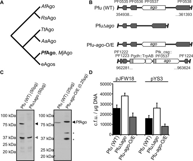
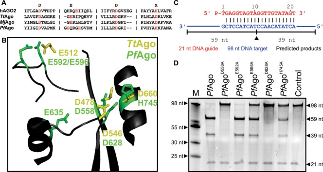
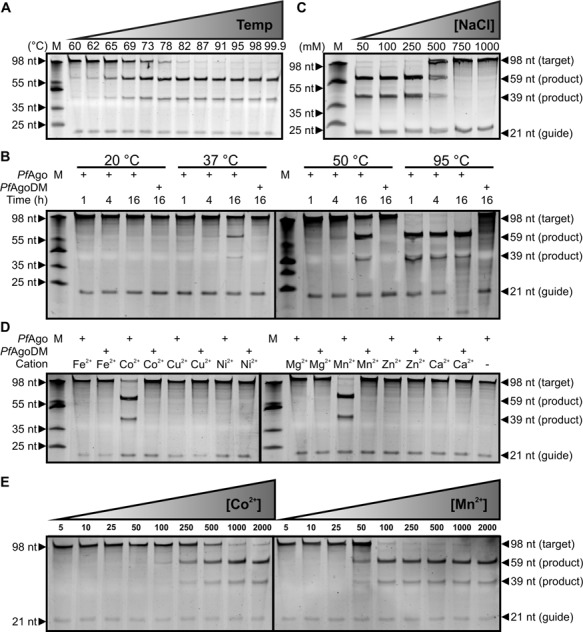
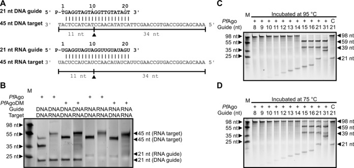
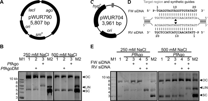
Functions of prokaryotic Argonautes (pAgo) have long remained elusive. Recently, Argonautes of the bacteria Rhodobacter sphaeroides and Thermus thermophilus were demonstrated to be involved in host defense. The Argonaute of the archaeon Pyrococcus furiosus (PfAgo) belongs to a different branch in the phylogenetic tree, which is most closely related to that of RNA interference-mediating eukaryotic Argonautes. Here we describe a functional and mechanistic characterization of PfAgo. Like the bacterial counterparts, archaeal PfAgo contributes to host defense by interfering with the uptake of plasmid DNA. PfAgo utilizes small 5'-phosphorylated DNA guides to cleave both single stranded and double stranded DNA targets, and does not utilize RNA as guide or target. Thus, with respect to function and specificity, the archaeal PfAgo resembles bacterial Argonautes much more than eukaryotic Argonautes. These findings demonstrate that the role of Argonautes is conserved through the bacterial and archaeal domains of life and suggests that eukaryotic Argonautes are derived from DNA-guided DNA-interfering host defense systems.


Figures

Figure 1.
Figure 1.
PfAgo interferes with plasmid transformation. (A) Schematic phylogenetic tree of Argonaute proteins, adapted from (3). AfAgo: Archaeoglobus fulgidus Ago. RsAgo: Rhodobacter sphaeroides Ago. TtAgo: Thermus thermophilus Ago. AaAgo: Aquifex aeolicus Ago. PfAgo: Pyrococcus furiosus Ago. MjAgo: Methanocaldococcus jannaschii Ago. eAgos: eukaryotic Agos. (B) Overview of ago gene loci of P. furiosus strains Pfu (wild-type), PfuΔago (knockout) and Pfu-ago-O/E (PfAgo overexpression strain). (C) Immunoblot analysis of PfAgo (indicated with a black triangle) content in Pfu and PfuΔago with Csa2 protein (indicated with a gray triangle) serving as the internal standard (left panel) or Pfu and Pfu-ago-O/E (right panel). The amount of lysate analyzed is indicated and the asterisk (*) denotes apparent breakdown products observed when PfAgo is overexpressed. (D) Plasmid transformation efficiencies of the P. furiosus strains. Error bars indicate standard deviations of biological triplicates.
Figure 2.
Figure 2.
DEDH catalytic site of PfAgo. (A) Sequence alignment of human AGO2 (hAGO2), TtAgo, MjAgo and PfAgo, adapted from (3). Only regions containing the DEDX catalytic residues (indicated in red) are shown. PfAgo catalytic residues E592 and E596 are colored orange. (B) TtAgo catalytic residues DEDD (yellow; PDB: 4N47) aligned to PfAgo catalytic site (black; PDB: 1Z25). Predicted catalytic residues of PfAgo are colored green. (C) Synthetic 21 nucleotide siDNA (red) and 98 nucleotide ssDNA target (blue) used for in vitro activity assays. The black triangle indicates the predicted cleavage site, black lines indicate the predicted 59 and 39 nucleotide cleavage products. (D) PfAgo and mutants were loaded with a 21 nucleotide long siDNA and were incubated with a 98 nucleotide ssDNA target in a 5:1:1 molar ratio (PfAgo:guide:target). Products were resolved on a 15% denaturing polyacrylamide gel. M: ssDNA marker. nt: nucleotide. The ‘Control’ sample contains no protein.
Figure 3.
Figure 3.
Effect of temperature, salt concentration and cation on PfAgo activity. PfAgo loaded with a 21 nt long siDNA was incubated with a 98 nt ssDNA target (see Figure 2C) in a 5:1:1 molar ratio (PfAgo:guide:target) under various conditions. Unless otherwise indicated, target cleavage took place at 95°C for 1 h, with 0.5 mM Mn2+ as cation. Nucleic acids are resolved on denaturing polyacrylamide gels. M: ssDNA marker. nt: nucleotide. (A) PfAgo activity is highest at temperatures between 90 and 99.9°C. (B) PfAgo shows activity at temperatures ≥37°C if incubation is extended. (C) NaCl concentrations ≥500 mM interfere with PfAgo activity. (D) PfAgo-guide complexes show Co2+ and Mn2+ mediated ssDNA target cleavage. (E) Mn2+ is preferred above Co2+ as cation for PfAgo-guide mediated ssDNA target cleavage.
Figure 4.
Figure 4.
PfAgo utilizes 15–31 nt long siDNAs for ssDNA target cleavage. PfAgo was incubated with various guides and targets in a 5:1:1 ratio (PfAgo:guide:target). Unless otherwise indicated, target cleavage took place at 75°C for 1 h, with 0.5 mM Mn2+ as cation. Nucleic acids are resolved on denaturing polyacrylamide gels. M: ssDNA marker. (A) Synthetic DNA and RNA guides (black) and targets (gray). The black triangle indicates the predicted cleavage site, black lines indicate the predicted 11 and 34 nt cleavage products. (B) PfAgo shows only DNA-guided DNA cleavage. (C and D) Both at 95 and 75°C, PfAgo-mediated target cleavage is facilitated by siDNAs which are at least 15 nt long. C: control reaction with PfAgoDM and 21 nt long siDNA.
Figure 5.
Figure 5.
Plasmid cleavage by PfAgo. (A) pWUR790 expression plasmid. (B) PfAgo expressed at 20°C and purified in absence of Mn2+ cleaves expression plasmid pWUR790. Agarose gels with plasmid targets incubated without protein (lane 1), with PfAgoDM (lane 2) and with PfAgo (lane 3). M1: 1-kb DNA ladder (New England Biolabs). M2: pWUR790 marker with open circular (OC), linearized (LIN) and supercoiled (SC) pWUR790. (C) pWUR704 target plasmid, target site indicated in gray. (D) Target region (gray) and FW and RV siDNA guides (black). Predicted cleavage sites are indicated with a black triangle. (E) Agarose gels with plasmid targets incubated with PfAgoDM (lane 1), with guide free PfAgo (lane 2) and with PfAgo loaded with FW siDNA, RV siDNA, or both (lane 3–5) in reaction buffer with 250 mM NaCl (left panel) or 500 mM NaCl (right panel). M1: 1 kb GeneRuler marker (Thermo Scientific). M2: pWUR704 marker with open circular (OC), linearized (LIN) and supercoiled (SC) pWUR704.

References

    1. Ketting R.F. The many faces of RNAi. Dev. Cell. 2011;20:148–161. - PubMed
    1. Meister G. Argonaute proteins: functional insights and emerging roles. Nat. Rev. Genet. 2013;14:447–459. - PubMed
    1. Swarts D.C., Makarova K., Wang Y., Nakanishi K., Ketting R.F., Koonin E.V., Patel D.J., van der Oost J. The evolutionary journey of Argonaute proteins. Nat. Struct. Mol. Biol. 2014;21:743–753. - PMC - PubMed
    1. Pratt A.J., MacRae I.J. The RNA-induced silencing complex: a versatile gene-silencing machine. J. Biol. Chem. 2009;284:17897–17901. - PMC - PubMed
    1. Shabalina S.A., Koonin E.V. Origins and evolution of eukaryotic RNA interference. Trends Ecol. Evol. 2008;23:578–587. - PMC - PubMed

Publication types

MeSH terms