. 2015 May 26;43(10):4923-36.
doi: 10.1093/nar/gkv422. Epub 2015 Apr 29.

A novel sigma factor reveals a unique regulon controlling cell-specific recombination in Mycoplasma genitalium

Affiliations

A novel sigma factor reveals a unique regulon controlling cell-specific recombination in Mycoplasma genitalium

Sergi Torres-Puig et al. Nucleic Acids Res. .

Abstract

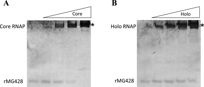
The Mycoplasma genitalium MG428 protein shows homology to members of the sigma-70 family of sigma factors. Herein, we found that MG428 activates transcription of recA, ruvA and ruvB as well as several genes with unknown function. Deletion of MG_428 or some of the up-regulated unknown genes led to severe recombination defects. Single cell analyses revealed that activation of the MG428-regulon is a rare event under laboratory growth conditions. A conserved sequence with sigma-70 promoter architecture (TTGTCA-N(18/19)-ATTWAT) was identified in the upstream region of all of the MG428-regulated genes or operons. Primer extension analyses demonstrated that transcription initiates immediately downstream of this sigma70-type promoter in a MG428-dependent manner. Furthermore, mutagenesis of the conserved -10 and -35 elements corroborated the requirement of these regions for promoter function. Therefore, a new mycoplasma promoter directs transcription of a unique recombination regulon. Additionally, MG428 was found to interact with the RNAP core enzyme, reinforcing the predicted role of this protein as an alternative sigma factor. Finally, our results indicate that MG428 contributes to the generation of genetic diversity in this model organism. Since recombination is an important mechanism to generate antigenic variation, MG428 emerges as a novel factor contributing to M. genitalium virulence.


Figures

Figure 1.
Figure 1.
Insertion points of the TnCatMG_428 MiniTnp in the M. genitalium genome. (A) Schematic representation depicting the genes preferentially targeted by TnCatMG_428 in the genome of a ΔMG_428 mutant. Genes disrupted by TnCatMG_428 are shown in blue. Black filled triangles represent the transposon insertion points. Arrows above the triangles indicate the orientation of the transposon insertion. (B) Schematic representation showing the presence of a truncated MG_428 ectopic copy in the genome of the Tn::MG390-1 and -2 mutants. The MG_428 coding region is highlighted in orange and the inverted repeats (IR) of the TnCatMG_428 MiniTnp are shown in green. The Tn::MG_281–4 mutant, carrying a full copy of the MG_428 gene, is also shown for comparison.
Figure 2.
Figure 2.
Analysis of protein expression by western blotting. (A) Immunoblot analysis of MG428 expression in the WT strain and several representative mutants. Lane 1, WT; lane 2, ΔMG_428; lane 3, Tn::MG_390-1; lane 4, Tn::MG_281-1; lane 5, Tn::recA-2; lane 6, Tn::MG_220-1; lane 7, Tn::MG_191-2 and lane 8, Tn::MG_192-1. (B) Immunoblot analysis of mCherry expression. Lane 1, WT; lane 2, Cat:Ch; lane 3, RecA:Ch; lane 4, MG_428:Ch; lane 5, ΔMG_428-RecA:Ch; lane 6, RecA:Ch-10; lane 7, RecA:Ch-22 and lane 8, RecA:Ch-35. Because the Cat-mCherry fusion is expressed at very high levels compared to the RecA-mCherry fusion, the amount of total protein loaded for the Cat:Ch mutant was reduced 20 times. HsdS protein was detected with a monoclonal antibody and used as a loading control.
Figure 3.
Figure 3.
Analysis of gene expression by qRT-PCR. Transcriptional analysis of selected M. genitalium genes in the WT and several mutant strains. Three independent biological repeats were performed and the respective fold-changes in gene expression are indicated with diamonds, squares and triangles. Mean fold-changes for each target gene are represented by color bars. Statistical significance of mean fold-changes above the cutoff (>2) was assessed with Student's t test. Statistically significant values (P < 0.05) are indicated with a red asterisk. Transcription of MG_220 (309 bp) in the Tn::MG_220 mutant could not be assessed by qRT-PCR due to the presence of a TnCatMG_428 MiniTnp insertion in this gene.
Figure 4.
Figure 4.
Analysis of MG428-RNA polymerase interaction. A fixed amount of soluble rMG428 protein (200 ng) was incubated with increased concentrations (0, 2.5, 5, 10 and 20 mM) of RNAP core enzyme (A) or RNAP holoenzyme (B). Mixtures were separated on native discontinuous 4–15% polyacrylamide gels and stained with colloidal Coomassie. Bands indicated with an asterisk (*) were cut off the gel and analyzed by LC–MS (see also Supplementary Table S6).
Figure 5.
Figure 5.
Identification of a conserved region with sigma-70 promoter architecture within the UR of the MG428-regulated genes. (A) Sequence logo generated with the UR of all the MG428-regulated genes or operons identified in this study. (B) Sequence logo generated with the UR of the respective M. pneumoniae homologs. The logo consists of stacks of letters, one stack for each position in the sequence analyzed. The overall height of each stack indicates the sequence conservation at that position (measured in bits). Nucleotides corresponding to the putative −35 and −10 promoter elements are boxed. Underlined and bolded nucleotides indicate experimentally determined TSSs (see also Figure 6). Due to the different length of the spacer region between the hexanucleotide promoter elements, one nucleotide gap was arbitrarily located immediately after the −35 box when necessary.
Figure 6.
Figure 6.
Identification of the TSSs of several MG428-regulated genes. Primer extension analysis of the MG_220, recA, MG_RS02200 and MG_414 genes in the WT strain and the Tn::recA-2 mutant. All electropherograms were generated with Peak Scanner v1.0 (Applied Biosystems) analysis software. Red peaks represent ROX size standards while blue peaks correspond to the primer extension products. Schematic representations of the genome regions analyzed are shown and the presence of the identified promoters indicated with blue arrows. The approximate location of the primers used in these analyses is also indicated by arrows.
Figure 7.
Figure 7.
Single cell analysis of Cat-, RecA- and MG428-mCherry expression by fluorescence microscopy. Each row contains a series of three images corresponding to the phase contrast, the Texas Red channel and the resulting overlay, respectively. Arrows indicate the presence of fluorescent cells expressing either RecA- or MG428-mCherry fusions. All pictures are shown at the same magnification.
Figure 8.
Figure 8.
Transformation efficiency by homologous recombination (TE-HR) of different M. genitalium strains. Bars show the averages and the standard deviations of at least three independent biological replicates for each strain. TE-HR of strains ΔMG_428, Tn::recA-2, Tn::ruvB, ΔMG_220 and ΔMG_RS02200 was below the limit of detection (<10−8). TE-HR that were found to be statistically different than that of the WT strain are indicated with an asterisk. Statistical significance was assessed with Student's t test (P < 0.05).
Figure 9.
Figure 9.
Whole-genome analysis of selected MG_428 complemented mutants by next generation sequencing. A scheme of the M. genitalium MgPa operon is shown along with its respective coverage data (in blue) and variant frequencies (in red). Discrete DNA repeats are boxed and numbered (R1 to R6). Genomic regions exhibiting a high rate of nucleotide sequence variation in the Tn::MG_192-1 mutant as compared to the WT strain are shadowed. Variations labeled with an asterisk (*) correspond to length polymorphisms of a trinucleotide repeat. A variation labeled with a pound sign (#) corresponds to the insertion point of the TnCatMG_428 MiniTnp in the Tn::MG_192-1 mutant (see Supplementary Table S5). Frequency of variants in the transposon insertion point is ∼50%. This result can be explained because reads of the positive strand show only sequence variants downstream of the transposon insertion point. Conversely, reads of the negative strand show only sequence variants upstream of the transposon insertion point. Since the graphic coverage shows combined reads from both the positive and the negative strand, this leads to a frequency of variation of about 50% in the regions flanking the transposon insertion site. Genomic regions exhibiting a high rate of nucleotide sequence variation in the Tn::MG_192-1 mutant as compared to the WT strain are shadowed.

References

    1. McGowin C.L., Anderson-Smits C. Mycoplasma genitalium: an emerging cause of sexually transmitted disease in women. PLoS Pathog. 2011;7:e1001324. - PMC - PubMed
    1. Gibson D.G., Glass J.I., Lartigue C., Noskov V.N., Chuang R.Y., Algire M.A., Benders G.A., Montague M.G., Ma L., Moodie M.M., et al. Creation of a bacterial cell controlled by a chemically synthesized genome. Science. 2010;329:52–56. - PubMed
    1. Gibson D.G., Benders G.A., Andrews-Pfannkoch C., Denisova E.A., Baden-Tillson H., Zaveri J., Stockwell T.B., Brownley A., Thomas D.W., Algire M.A., et al. Complete chemical synthesis, assembly, and cloning of a Mycoplasma genitalium genome. Science. 2008;319:1215–1220. - PubMed
    1. Karr J.R., Sanghvi J.C., Macklin D.N., Gutschow M.V., Jacobs J.M., Bolival B. Jr, Assad-Garcia N., Glass J.I., Covert M.W. A whole-cell computational model predicts phenotype from genotype. Cell. 2012;150:389–401. - PMC - PubMed
    1. Zhang W., Baseman J.B. Transcriptional regulation of MG_149, an osmoinducible lipoprotein gene from Mycoplasma genitalium. Mol. Microbiol. 2011;81:327–339. - PMC - PubMed

Publication types