. 2015 May 26;43(10):5182-93.
doi: 10.1093/nar/gkv387. Epub 2015 Apr 29.

The ribonuclease DIS3 promotes let-7 miRNA maturation by degrading the pluripotency factor LIN28B mRNA

Affiliations

The ribonuclease DIS3 promotes let-7 miRNA maturation by degrading the pluripotency factor LIN28B mRNA

Simona Segalla et al. Nucleic Acids Res. .

Abstract

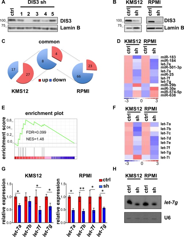
Multiple myeloma, the second most frequent hematologic tumor after lymphomas, is an incurable cancer. Recent sequencing efforts have identified the ribonuclease DIS3 as one of the most frequently mutated genes in this disease. DIS3 represents the catalytic subunit of the exosome, a macromolecular complex central to the processing, maturation and surveillance of various RNAs. miRNAs are an evolutionarily conserved class of small noncoding RNAs, regulating gene expression at post-transcriptional level. Ribonucleases, including Drosha, Dicer and XRN2, are involved in the processing and stability of miRNAs. However, the role of DIS3 on the regulation of miRNAs remains largely unknown. Here we found that DIS3 regulates the levels of the tumor suppressor let-7 miRNAs without affecting other miRNA families. DIS3 facilitates the maturation of let-7 miRNAs by reducing in the cytoplasm the RNA stability of the pluripotency factor LIN28B, a inhibitor of let-7 processing. DIS3 inactivation, through the increase of LIN28B and the reduction of mature let-7, enhances the translation of let-7 targets such as MYC and RAS leading to enhanced tumorigenesis. Our study establishes that the ribonuclease DIS3, targeting LIN28B, sustains the maturation of let-7 miRNAs and suggests the increased translation of critical oncogenes as one of the biological outcomes of DIS3 inactivation.


Figures

Figure 1.
Figure 1.
DIS3 knockdown in MM cells selectively affects let-7 miRNA family. (A) Western blot to test the efficiency of five different shRNAs to silence DIS3 in HEK-293T cell line. (B) Western blot analysis of DIS3 expression in MM cell lines, KMS12-BM (KMS12) AND RPMI-8226 (RPMI), infected with a scrambled shRNA (ctrl) or DIS3 shRNA4 (shRNA). Lamin B was used as loading control. (C) Pie charts summarizing the number of miRNAs deregulated >1.5-fold in KMS12 and RPMI, and in both cell lines after DIS3 knockdown. (D) Heat map showing the expression levels of miRNA varying >1.5-fold in both KMS12 and RPMI upon DIS3 silencing. The color scale bar in the heat map represents the relative miRNA expression with red representing up-regulation and blue representing down-regulation. (E) GSEA ES plot showing the enrichment of let-7 miRNA family gene set after DIS3 silencing. (F) Heat map of let-7 miRNA family levels in KMS12 and RPMI cell lines after infection with a scrambled shRNA (ctrl) or DIS3-specific shRNA4 (sh). (G) Expression of the let-7 miRNA members (a, b, f, g) assayed by qRT-PCR in KMS12 and RPMI cell lines infected with scrambled (ctrl) or DIS3-specific shRNA4 (sh), 72 h after infection. Results are normalized over RNU6B. Bars represent SDs (n = 2 indipendent experiments). *P < 0.05; **P < 0.005 using two-tailed Student's t test. (H) Northern blot analysis of endogenous let-7g in KMS12 and RPMI cell lines infected with scrambled (ctrl) or DIS3-specific shRNA4 (sh), 72 h after infection. RNU6B was used as loading control.
Figure 2.
Figure 2.
let-7 levels after DIS3 knockdown in human and mouse cell lines. (A) Western blot analysis of DIS3 expression in human U2OS and HEK293T cells, and in mouse NIH3T3 cells, 72 h after infection with scrambled (ctrl) and human or mouse DIS3-specific shRNAs (sh). Results are normalized over loading control Lamin B and RAN. (B) Expression of the let-7 miRNA members was assayed by qRT-PCR. Results are normalized over RNU6B and miR-16 respectively for human and mouse cells. Bars represent SDs (n = 2 indipendent experiments). *P < 0.05 using two-tailed Student's t test.
Figure 3.
Figure 3.
DIS3 silencing increases MYC and RAS proteins and induces transformation. (A) Representative blot and quantification of DIS3, MYC and RAS proteins in RPMI, KMS12 and NIH3T3 cells, 72 h after infection with scrambled (ctrl) or human and mouse DIS3-specific shRNAs (sh). Results are normalized over loading control lamin B and RAN, respectively for MM cells and NIH3T3 cells. The error bars represent SD of two independent experiments. (B) MYC and RAS mRNA levels normalized over GAPDH in the same cells of panel (A). Bars represent SDs (n = 2 indipendent experiments). *P < 0.05; **P < 0.005. (C) U2OS infected with scrambled (ctrl) or DIS3-specific (sh) shRNAs were treated with 100 μg/ml cycloheximide for the time indicated and lysates were immunoblotted for DIS3 and MYC. Lamin B represents a loading control. (D) Western blot and quantification of levels for endogenous DIS3 and MYC in U2OS cells knocked-down with a scrambled shRNA (ctrl) or a DIS3 specific shRNA (shRNA4, sh), 48 h after transfection with let-7a (+) or control RNA (–) mimics. Lamin B was used as loading control. (E) Western blot of DIS3 levels and focus formation assay of NIH3T3 cells infected with scrambled shRNA (ctrl) or with murine DIS3 shRNA4 (sh). Colonies were counted from three independent platings. The error bars represent SD. **P < 0.005 using two-tailed Student's t test. RAN represents the loading control.
Figure 4.
Figure 4.
DIS3 affects let-7 processing regulator LIN28B. (A) Measurement by qRT-PCR of the pri-miRNAs let-7-a/f/d (left panel) and mature miRNA let-7-a (right panel) in RPMI, KMS12 and U2OS cells, 72 h after infection with scrambled (ctrl) or DIS3-specific shRNA (sh). pri-miRNAs let-7-a/f/d expression data were normalized over GAPDH. let-7-a expression data were normalized over RNU6B. Bars represent SDs (n = 2 indipendent experiments). *P < 0.05; **P < 0.005 using two-tailed Student's t test. (B) LIN28A and LIN28B mRNA levels assessed by qRT-PCR with respect to GAPDH expression 72 h after infection. Bars represent SDs (n = 2 indipendent experiments). **P < 0.005 using two-tailed Student's t test. (C) Blot (left panel) and quantification (right panel) representative for two experiments showing DIS3 and LIN28B protein levels in RPMI, KMS12 and U2OS 72 h after infection with shRNA targeting DIS3. Lamin B was used as loading control.
Figure 5.
Figure 5.
DIS3 controls let-7 through LIN28B. LIN28B mRNA (left panel) and let-7-a and let-7-g (right panel) levels of one representative experiment in which RPMI cells infected with a scrambled shRNA (ctrl) or with a DIS3 shRNA4 (DIS3 sh) underwent, after 3 days, a second infection with a scrambled shRNA (ctrl) or with a LIN28B shRNA. LIN28B and let-7 levels were measured 4 days after the second infection and normalized over GAPDH and RNU6B respectively. Bars represent SDs (n = 2 replicas of one representative experiment).
Figure 6.
Figure 6.
DIS3 affects RNA stability of LIN28B RNA. U2OS cells were infected with a scrambled shRNA (control sh) or a DIS3 shRNA (shRNA4) (A) or were transfected with a control siRNA (ctrl) or a siRNA against SKI (B) or TRAMP (C). 3 days after infection and selection or 48 h after transfection, cells were treated with DRB (100 μg/ml) to block RNA synthesis. The stability of LIN28B and GAPDH total mRNA was measured by qRT-PCR at 0, 2, 3 and 4 h after treatment and expressed as relative abundance with respect to mRNA level at 0 h. Bars represent SDs (n = 2 indipendent experiments in A; n = 2 replicas of one representative experiment in B and C). *P < 0.05 using two-tailed Student's t test.
Figure 7.
Figure 7.
Proposed model on the role of DIS3 in regulating LIN28B, let-7 and MYC and RAS.

References

    1. Schneider C., Leung E., Brown J., Tollervey D. The N-terminal PIN domain of the exosome subunit Rrp44 harbors endonuclease activity and tethers Rrp44 to the yeast core exosome. Nucleic Acids Res. 2009;37:1127–1140. - PMC - PubMed
    1. Mitchell P., Petfalski E., Shevchenko A., Mann M., Tollervey D. The exosome: a conserved eukaryotic RNA processing complex containing multiple 3′–>5′ exoribonucleases. Cell. 1997;91:457–466. - PubMed
    1. Houseley J., LaCava J., Tollervey D. RNA-quality control by the exosome. Nat. Rev. Mol. Cell Biol. 2006;7:529–539. - PubMed
    1. Liu Q., Greimann J.C., Lima C.D. Reconstitution, activities, and structure of the eukaryotic RNA exosome. Cell. 2006;127:1223–1237. - PubMed
    1. Hernandez H., Dziembowski A., Taverner T., Seraphin B., Robinson C.V. Subunit architecture of multimeric complexes isolated directly from cells. EMBO Rep. 2006;7:605–610. - PMC - PubMed

Publication types