. 2015;7(5):506-17.
doi: 10.1159/000381213. Epub 2015 Apr 25.

Collagen VI Is Upregulated in COPD and Serves Both as an Adhesive Target and a Bactericidal Barrier for Moraxella catarrhalis

Affiliations

Collagen VI Is Upregulated in COPD and Serves Both as an Adhesive Target and a Bactericidal Barrier for Moraxella catarrhalis

Suado M Abdillahi et al. J Innate Immun. 2015.

Abstract

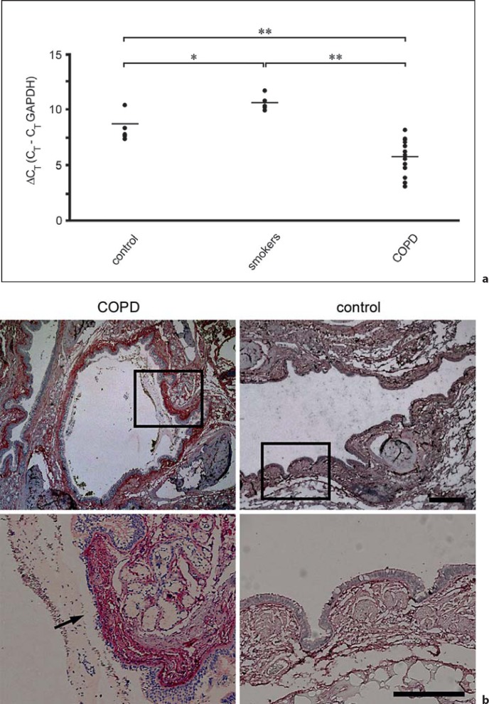
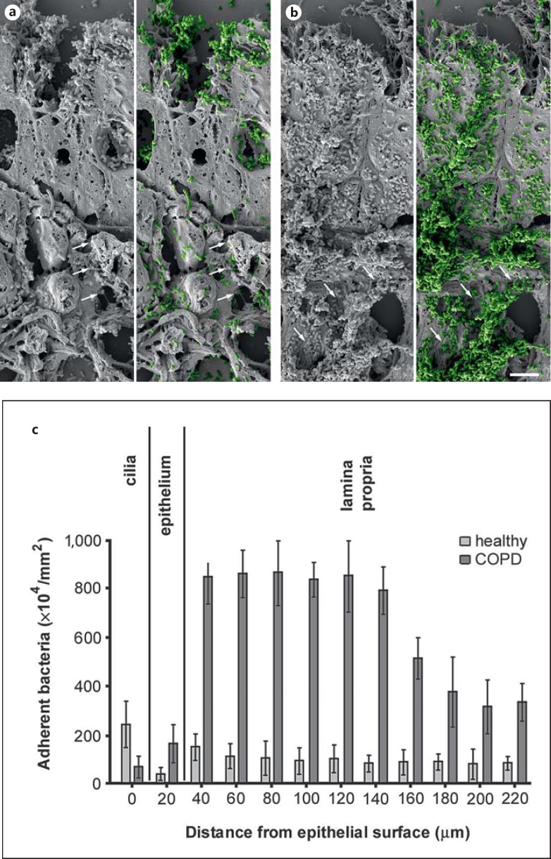
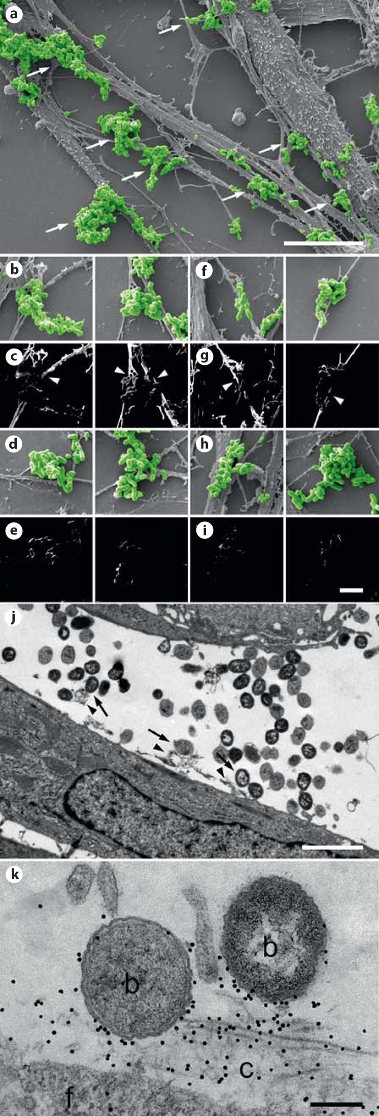
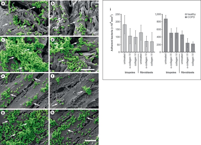
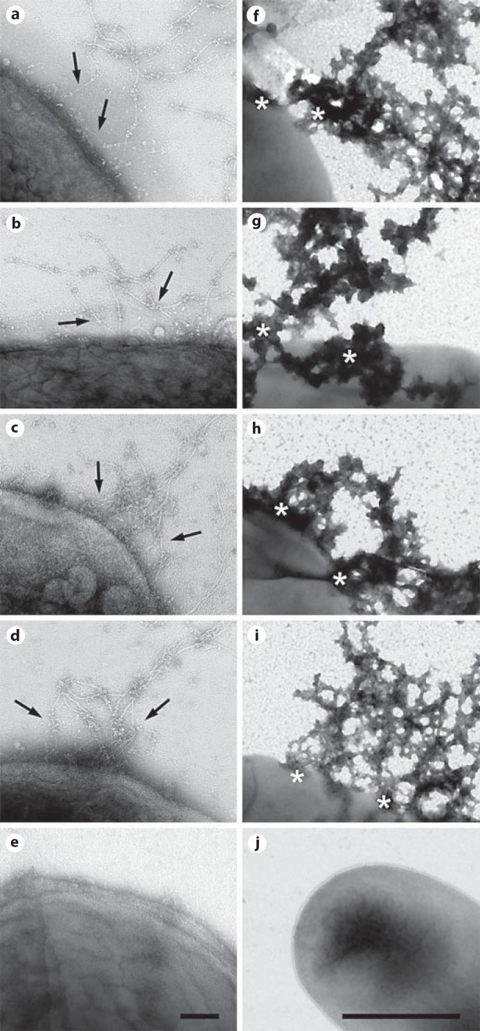
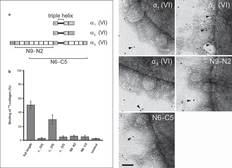
Moraxella catarrhalis is a Gram-negative human mucosal commensal and pathogen. It is a common cause of exacerbation in chronic obstructive pulmonary disease (COPD). During the process of infection, host colonization correlates with recognition of host molecular patterns. Importantly, in COPD patients with compromised epithelial integrity the underlying extracellular matrix is exposed and provides potential adhesive targets. Collagen VI is a ubiquitous fibrillar component in the airway mucosa and has been attributed both adhesive and killing properties against Gram-positive bacteria. However, less is known regarding Gram-negative microorganisms. Therefore, in the present study, the interaction of M. catarrhalis with collagen VI was characterized. We found that collagen VI is upregulated in the airways of COPD patients and exposed upon epithelial desquamation. Ex vivo, we inoculated airway biopsies and fibroblasts from COPD patients with M. catarrhalis. The bacteria specifically adhered to collagen VI-containing matrix fibrils. In vitro, purified collagen VI microfibrils bound to bacterial surface structures. The primary adhesion target was mapped to the collagen VI α2-chain. Upon exposure to collagen VI, bacteria were killed by membrane destabilization in physiological conditions. These previously unknown properties of collagen VI provide novel insights into the extracellular matrix innate immunity by quickly entrapping and killing pathogen intruders.


Figures

Fig. 1
Fig. 1
Collagen VI is upregulated in COPD airways and exposed in regions of epithelial damage. a RT-PCR of collagen VI expression in lung tissues from healthy individuals, smokers and COPD patients. Relative gene expression was normalized to the housekeeping gene GAPDH and is given as ΔCT value, where higher values correspond to lower gene expression levels. Data sets were assessed by Student's t test for paired data. Differences were considered statistically significant at p ≤ 0.05. Significance levels were set at * p ≤ 0.05 and ** p ≤ 0.01. b Immunohistochemistry of airway biopsies obtained from patients with COPD (left panel) and from healthy control individuals (right panel). Collagen VI is widely distributed in the subepithelial lamina propria (control panel). It is overexpressed in COPD, especially in a zone 120 ± 40 μm wide distant to the basement membrane (COPD panel). Regions of epithelial damage and exposure of collagen VI in the underlying connective tissue are frequently observed (arrow). The scale bars represent 500 μm (upper panel) and 250 μm (lower panel).
Fig. 2
Fig. 2
Colonization of M. catarrhalis in COPD lung tissues ex vivo. Paraffin sections of lung biopsies from healthy individuals and COPD patients were inoculated with M. catarrhalis. In COPD, large amounts of bacteria adhered to the airway mucosa (b) as compared to only moderate binding to tissues from healthy subjects (a). M. catarrhalis is highlighted in green color in a and b. Frequently bacteria were bound to fibrillar extracellular structures (arrows). The scale bar represents 5 μm. c Quantification of adherent bacteria found in different compartments of the airway mucosa.
Fig. 3
Fig. 3
M. catarrhalis bind to collagen VI-containing extracellular matrix fibrils. Human lung fibroblasts from patients with severe COPD (GOLD stage IV; a, c) were allowed to deposit a dense fibrillar matrix. The specimens were incubated with M. catarrhalis and prepared for immunolocalization and scanning electron microscopy. The bacteria were frequently observed to adhere to extracellular collagen fibrils (arrows). These fibrils contained collagen VI as shown by immunodetection with collagen VI antibodies (b, c, arrowheads). Similar observations were made with collagen fibrils in airway biopsies described in figure 2 (f, g). Control specimens treated with secondary gold conjugate alone did not exhibit any fibrillar staining (d, e, fibroblasts; h, i, airway biopsies). The scale bars represent 10 μm (a) and 2.5 μm (b-i). j, k Ultrathin sections of fibroblasts incubated with M. catarrhalis bacteria (arrows) adhering to the extracellular collagen matrix (arrowheads). k Bacteria are frequently colocalized with collagen VI in the extracellular collagen matrix as visualized by collagen VI antibodies conjugated with 5-nm colloidal gold. f = Fibroblast; b = bacteria; c = extracellular collagen matrix. The scale bars represent 1 μm.
Fig. 4
Fig. 4
Blocking of M. catarrhalis adherence to the airway mucosa and human pulmonary fibroblasts. Scanning electron micrographs of paraffin sections of airway biopsies (a-d) or pulmonary fibroblasts (e-h) from healthy individuals (a, b, biopsies; e, f, fibroblasts) and COPD patients (c, d, biopsies; g, h, fibroblasts) inoculated with M. catarrhalis bacteria in the absence (a, c, e, g) or presence (b, d, f, h) of α-collagen VI antibodies. The scale bars represent 5 μm. i Quantitative evaluation of adherent bacteria.
Fig. 5
Fig. 5
Collagen VI microfibrils adhere to M. catarrhalis and other pulmonary pathogens, followed by killing. Collagen VI microfibrils were purified from bovine cornea and allowed to react with M. catarrhalis (a), nontypeable H. influenzae (b), S. pneumoniae (c) and P. aeruginosa (d). Negative staining and transmission electron microscopy visualizes primary adhesion of collagen VI networks (arrows) to the bacterial surfaces (a-d), followed by killing within 1 h (f-i). Asterisks denote areas of membrane destabilization and exudation of intracellular content (f-i). F. magna was used as a control and did neither recruit collagen VI (e) nor exhibit killing (j). The scale bars represent 100 nm (a-e) and 1 μm (f-j).
Fig. 6
Fig. 6
Collagen VI microfibrils induce killing of pulmonary pathogens by membrane destabilization. Bacteria were incubated with 1 mM collagen VI for 2 h at 37°C. a Numbers of bacterial colonies after incubation with collagen VI were determined in viable count assays. Incubation in buffer alone (control) or LL-37 served as negative and positive controls, respectively. The dots represent individual experiments with the respective strains. F. magna were not killed and served as a negative control. Values are expressed as percent bacterial survival. b-g Negative staining and transmission electron microscopy of M. catarrhalis (b, d, f) and P. aeruginosa (c, e, g) bacteria. Upon incubation with collagen VI, large-scale membrane disruption results in destruction of the pathogens (d, e) as compared to controls incubated with TBS alone (b, c). When the bacteria are beginning to die, blebbing of membrane vesicles (arrowheads) is frequently observed (f, g). Similar observations were made for the killing of H. influenzae and S. pneumoniae (not shown). The scale bars represent 1 μm (b-e) and 0.5 μm (f, g).
Fig. 7
Fig. 7
The collagen VI α2-chain interacts specifically with M. catarrhalis. Recombinant collagen VI α-chains (α1, α2 and α3) and α3-chain fragments (N9-N2 and N6-C5; schematic overview in a) were labeled with 125I (b) or colloidal gold (c) and allowed to interact with M. catarrhalis. Only full-length collagen VI and the collagen VI α2-chain were specifically associated with bacteria (b, c, arrowheads). The scale bar represents 50 nm.

References

    1. Karalus R, Campagnari A. Moraxella catar rhalis: a review of an important human mucosal pathogen. Microbes Infect. 2000;2:547–559. - PubMed
    1. Verduin CM, Hol C, Fleer A, van Dijk H, van Belkum A. Moraxella catarrhalis: from emerging to established pathogen. Clin Microbiol Rev. 2002;15:125–144. - PMC - PubMed
    1. Aebi C. Moraxella catarrhalis - pathogen or commensal? Adv Exp Med Biol. 2011;697:107–116. - PubMed
    1. Stenfors LE, Raisanen S. Interaction between Streptococcus pneumoniae and Branhamella catarrhalis obtained from double-colonized, healthy nasopharynx and double-infected, diseased middle-ear cavity. Scand J Infect Dis. 1989;21:397–401. - PubMed
    1. Murphy TF, Parameswaran GI. Moraxella catarrhalis, a human respiratory tract pathogen. Clin Infect Dis. 2009;49:124–131. - PubMed

Publication types

MeSH terms

Substances