. 2015 Sep 15;21(18):4201-11.
doi: 10.1158/1078-0432.CCR-14-3049. Epub 2015 Apr 29.

Whole Exome Sequencing Reveals the Order of Genetic Changes during Malignant Transformation and Metastasis in a Single Patient with NF1-plexiform Neurofibroma

Affiliations

Whole Exome Sequencing Reveals the Order of Genetic Changes during Malignant Transformation and Metastasis in a Single Patient with NF1-plexiform Neurofibroma

Angela C Hirbe et al. Clin Cancer Res. .

Abstract

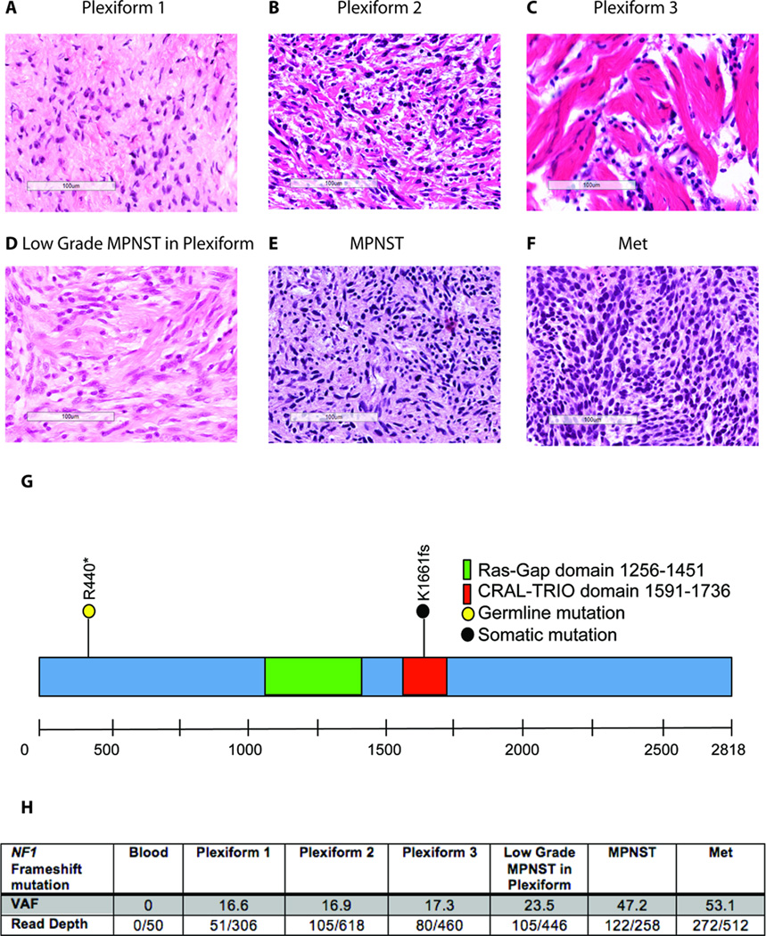
Purpose: Malignant peripheral nerve sheath tumors (MPNST) occur at increased frequency in individuals with neurofibromatosis type 1 (NF1), where they likely arise from benign plexiform neurofibroma precursors. While previous studies have used a variety of discovery approaches to discover genes associated with MPNST pathogenesis, it is currently unclear what molecular events are associated with the evolution of MPNST from plexiform neurofibroma.

Experimental design: Whole-exome sequencing was performed on biopsy materials representing plexiform neurofibroma (n = 3), MPNST, and metastasis from a single individual with NF1 over a 14-year period. Additional validation cases were used to assess candidate genes involved in malignant progression, while a murine MPNST model was used for functional analysis.

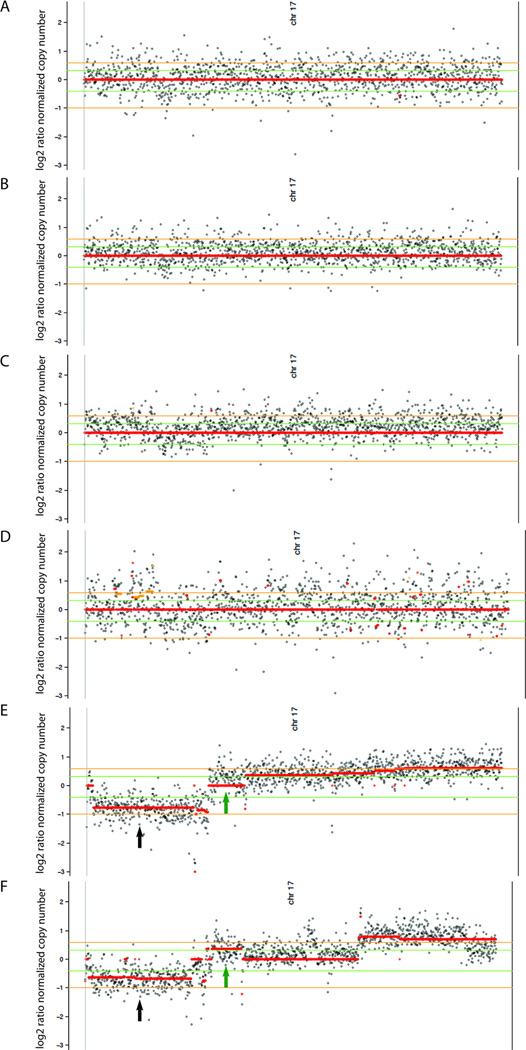
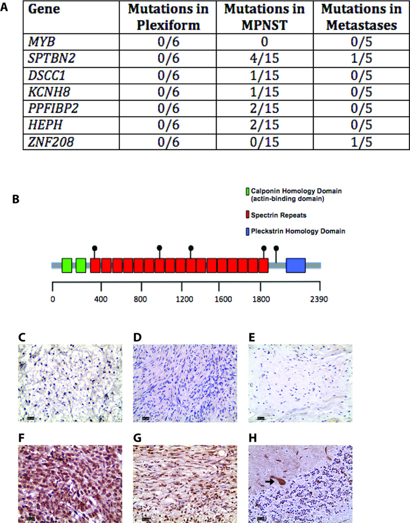
Results: There was an increasing proportion of cells with a somatic NF1 gene mutation as the tumors progressed from benign to malignant, suggesting a clonal process in MPNST development. Copy number variations, including loss of one copy of the TP53 gene, were identified in the primary tumor and the metastatic lesion, but not in benign precursor lesions. A limited number of genes with nonsynonymous somatic mutations (βIII-spectrin and ZNF208) were discovered, several of which were validated in additional primary and metastatic MPNST samples. Finally, increased βIII-spectrin expression was observed in the majority of MPNSTs, and shRNA-mediated knockdown reduced murine MPNST growth in vivo.

Conclusions: Collectively, the ability to track the molecular evolution of MPNST in a single individual with NF1 offers new insights into the sequence of genetic events important for disease pathogenesis and progression for future mechanistic study.


Conflict of interest statement

Conflicts of Interest: The authors have no conflicts to disclose.

Figures

Figure 1
Figure 1
Identification of the germline NF1 gene mutation in the discovery patient and the somatic NF1 gene mutation in tumor samples from this individual. Representative images of the Plexiform 1 (A), Plexiform 2 (B), Plexiform 3 (C), Low-Grade MPNST within Plexiform (D), primary high-grade MPNST (E), and metastatic MPNST (F). All images are at 20× magnification. Scale bars, 100µm. Diagram of the NF1 gene depicting the germline and somatic mutations identified in the discovery patient (G). Variant allele frequencies (VAF) with read depths for the somatic NF1 gene mutation in each tumor sample from the discovery patient (H).
Figure 2
Figure 2
Copy number plots depicting no alteration at chromosome 17 in Plexiform 1(A), Plexiform 2 (B), Plexiform 3 (C), and Low-Grade MPNST within Plexiform (D). In contrast, loss of one copy of the area of chromosome 17 encompassing the TP53 gene was observed in the primary (E) and metastatic (F) MPNST lesions. The area localizing to TP53 is indicated with a black arrow. The green arrow denotes the area of chromosome 17 amplified in the MPNST and metastatic tumor. The x-axis is the log2 ratio of the normalized copy number. All samples are normalized to a 0 copy number state. Positive numbers denote amplification, while negative numbers denote deletions. Red lines represent areas that the software calls constant copy number variation. Green lines represent one gain or loss in half of the cells. Orange lines represent one gain or loss in all of the cells.
Figure 3
Figure 3
Genes mutated in the MPNST validation set (A). A schematic representation of the βIII-spectrin protein with the location of the missense mutations denoted. Mutations were also identified in the splice region predicting exon skipping and in the 5’ untranslated region (not pictured). Positive βIII-spectrin immunostaining was not found in neurofibromas (C), schwannomas (D), or normal peripheral nerve (E), but was observed in the majority of MPNSTs with SPTBN2 mutations (F) as well as in MPNSTs without SPTBN2 mutations (G). (H) Normal cerebellum was included as an internal control for positive βIII-spectrin immunostaining. Arrow denotes the βIII-spectrin-immunoreactive Purkinje cells.
Figure 4
Figure 4
βIII-spectrin knockdown attenuates MPNST growth in vivo. (A) Western blotting reveals decreased βIII-spectrin expression in shSptbn2-infected JW18.2 cells relative to control shLacZ-infected cells. α-tubulin was included as an internal protein loading control. (B) In vitro cell proliferation was assessed by BLI at 24, 48, and 96 hours post-plating (p=0.276). (C) In vivo tumor growth was assessed by BLI at 5, 9, 12, and 16 days post-injection. Photon flux was quantitated in a fixed region of interest (ROI) over the subcutaneously implanted tumor (P<0.001; n=5 mice/group). (D) Representative images of mice injected with shSptbn2-infected cells relative to those injected with control shLacZ-infected cells are shown. (E) Gross images of the dissected tumors are shown.

Similar articles

Cited by

References

    1. Arun D, Gutmann DH. Recent advances in neurofibromatosis type 1. Current opinion in neurology. 2004;17:101–105. - PubMed
    1. Hirbe AC, Gutmann DH. Neurofibromatosis type 1: a multidisciplinary approach to care. The Lancet Neurology. 2014;13:834–843. - PubMed
    1. Ferner RE, Huson SM, Thomas N, Moss C, Willshaw H, Evans DG, et al. Guidelines for the diagnosis and management of individuals with neurofibromatosis 1. Journal of medical genetics. 2007;44:81–88. - PMC - PubMed
    1. Upadhyaya M, Spurlock G, Monem B, Thomas N, Friedrich RE, Kluwe L, et al. Germline and somatic NF1 gene mutations in plexiform neurofibromas. Human mutation. 2008;29:E103–E111. - PubMed
    1. Rasmussen SA, Overman J, Thomson SA, Colman SD, Abernathy CR, Trimpert RE, et al. Chromosome 17 loss-of-heterozygosity studies in benign and malignant tumors in neurofibromatosis type 1. Genes, chromosomes & cancer. 2000;28:425–431. - PubMed

Publication types