. 2015 May 15;142(10):1869-78.
doi: 10.1242/dev.115329. Epub 2015 Apr 29.

The transmembrane protein Crumbs displays complex dynamics during follicular morphogenesis and is regulated competitively by Moesin and aPKC

Affiliations

The transmembrane protein Crumbs displays complex dynamics during follicular morphogenesis and is regulated competitively by Moesin and aPKC

Kristin M Sherrard et al. Development. .

Erratum in

Abstract

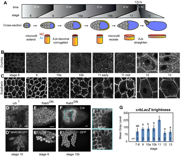
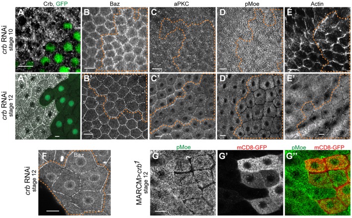
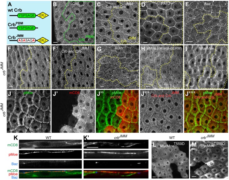
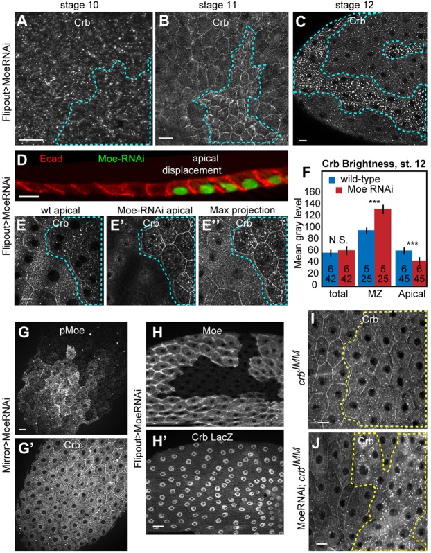
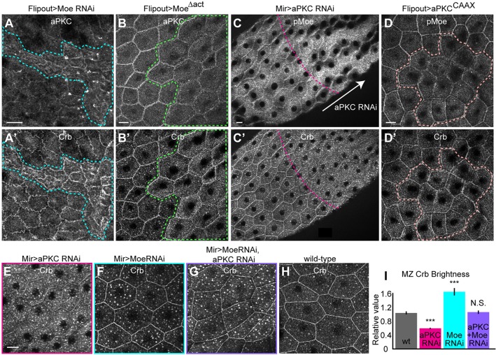
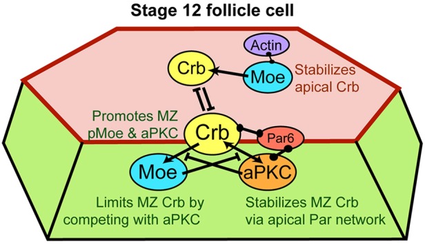
The transmembrane protein Crumbs (Crb) functions in apical polarity and epithelial integrity. To better understand its role in epithelial morphogenesis, we examined Crb localization and dynamics in the late follicular epithelium of Drosophila. Crb was unexpectedly dynamic during middle-to-late stages of egg chamber development, being lost from the marginal zone (MZ) in stage 9 before abruptly returning at the end of stage 10b, then undergoing a pulse of endocytosis in stage 12. The reappearance of MZ Crb is necessary to maintain an intact adherens junction and MZ. Although Crb has been proposed to interact through its juxtamembrane domain with Moesin (Moe), a FERM domain protein that regulates the cortical actin cytoskeleton, the functional significance of this interaction is poorly understood. We found that whereas the Crb juxtamembrane domain was not required for adherens junction integrity, it was necessary for MZ localization of Moe, aPKC and F-actin. Furthermore, Moe and aPKC functioned antagonistically, suggesting that Moe limits Crb levels by reducing its interactions with the apical Par network. Additionally, Moe mutant cells lost Crb from the apical membrane and accumulated excess Crb at the MZ, suggesting that Moe regulates Crb distribution at the membrane. Together, these studies reveal reciprocal interactions between Crb, Moe and aPKC during cellular morphogenesis.

Keywords: Crumbs; ERM proteins; F-actin; Par3/Bazooka; Squamous morphogenesis; aPKC.


Figures

Fig. 1.
Fig. 1.
Crumbs localization shifts in conjunction with squamous morphogenesis in FCs. (A) Major events during oogenesis stages 8-12: main body FCs (yellow) become columnar in stages 8-10a, then cuboidal and finally squamous, flattening in 20 min in stage 11 as the nurse cells (gray) dump their contents into the oocyte (blue). (B) Crb was lost from the MZ in stages 9-10, reappearing abruptly at the beginning of stage 11. Images are projections of four confocal sections, 0.3 µm apart, spanning the MZ and apical surface, imaged and processed with the same settings. Throughout the article, maximal projections encompassing the apical junctions and apices are shown, except where noted. (C) Ecad became highly corrugated in stages 9-11, straightening in 11-12; single sections were imaged and processed with the same settings. (D) Crb was present on follicle cell apices in stage 10, not merely on the oocyte surface, as shown by crb null mitotic-follicle cell clones positively labeled with MARCM. (E-F‴) Inhibiting endocytosis with flip-out clones expressing GFP and dominant-negative Rab5 (Rab5DN) did not affect Crb levels in stage 9 (E,E′), in contrast to stage 10 and later, in which it caused a strong buildup of Crb (F,F′). Close-up views of Crb staining in Rab5DN show apical buildup (F″) and lateral accumulation (F‴). (G) Crb transcription was upregulated in stages 9-11 relative to other stages, shown by mean gray levels of β-galactosidase in crb lacZ (crbM1M2) egg chambers. Scale bars: 10 µm (in C-E), 20 µm (in F,G). Error bars in G are s.e.m., n shows number of egg chambers measured (10-20 cells/egg chamber). Groups labeled a-c delineate those significantly different in unpaired, two-tailed t-tests, to P<0.01.
Fig. 2.
Fig. 2.
Crumbs is required to localize MZ and AJ components only after stage 10. (A) Driving crb RNAi in flip-out clones, positively labeled with GFP, caused strong loss of Crb, which, however, did not prevent cell expansion in stage 12 (A′). (B,C,D,E) crb depletion did not affect AJ or MZ proteins in stage 10, when Crb is normally absent from the MZ. However, crb RNAi caused gaps in Baz and loss at the MZ by stage 12 (B′,C′,D′,E′). (F) High-magnification view of Baz staining in crb RNAi cells. (G-G″) Single-section staining of pMoe and mCD8-RFP in crb null clones show that absence of pMoe staining does not correspond to a gap between cells. Dashed lines show crb RNAi flip-out clones (on the right hand in all images); in all figures, the outlines are placed slightly outside the clone MZ to show border with wild-type cells. Scale bars: 10 µm.
Fig. 3.
Fig. 3.
Crumbs JM domain is necessary for localization of MZ proteins but not AJs. (A) Diagram of knock-in alleles deleting each of the two conserved cytoplasmic domains of Crb; extracellular domains were intact in both alleles. (B) crbPBM, lacking the ERLI domain that binds to the apical Par proteins and Patj-Sdt complex, was unable to localize stably to the membrane and is present primarily in vesicles. (C) By contrast, the crbJMM allele could localize to the MZ and was endocytosed at the same level as in wt cells. In crbJMM clones, Patj (D) was normally localized, and Baz (E) was only slightly affected. However, MZ aPKC (F), F-actin (G) and pMoe (H, stained using a different fix and antibody from that in J and in Fig. 2D′,F; see Materials and Methods) were highly reduced in crbJMM clones, although total Moe level was normal (I). (J-J‴′) Single-section co-staining of pMoe, mCD8-GFP and Crb show that membrane is present within the gap in pMoe staining. (K,K′) Cross-sections of wild-type cells and crbJMM clones co-stained for pMoe, Baz and mCD8. (L,M) Expressing the constitutively phosphomimetic Myc-MoeT559D in crbJMM clones showed that, relative to wild-type controls, the crbJMM clones were still unable to localize MoeT559D to the MZ. Dashed lines show crb knock-in allele clones (on the right hand in all images). Scale bars: 10 µm. All images show stage 12-early 13 egg chambers.
Fig. 4.
Fig. 4.
In Moe-depleted cells after stage 10, Crb was excessively endocytosed, and accumulated at the MZ. Moe depletion did not affect Crb localization through stage 10 (A), but, beginning in stage 11 (B), caused accumulation of Crb at the MZ. (C) At the time wt cells endocytose Crb, Moe-depleted cells had larger and brighter Crb-stained vesicles. (D) Cross-section of stage 10 shows that Moe RNAi cells extend further apically than wt cells. In consequence, Moe-depleted cells are offset from wt cells, as sections E and E′ (1.2 µm apart) show; maximal projection in E″. Confocal projections shown elsewhere span the MZ to apical of wt and Moe-depleted cells. (F) Although Moe depletion did not change total Crb levels, it was associated with significantly more MZ Crb and less apical Crb. (G,G′) Depleting Moe with the weaker driver MirrorGal4, Gal80ts elevated MZ Crb without decreasing cell perimeter. (H,H′) Loss of Moe did not affect expression of the crb-lacZ transcriptional reporter. (I,J) In comparison to the crbJMM mutant alone (I), depleting Moe in a crbJMM mutant background reduced accumulation of Crb in the MZ. All egg chambers are stage 12, except where noted. Dashed lines show Moe RNAi flip-out clones or Moe null clones. Scale bars: 10 µm. Error bars show s.e.m.; *** denotes significant difference of P<0.001 by paired (values measured within the same egg chamber), two-tailed t-tests.
Fig. 5.
Fig. 5.
aPKC stabilized Crb at the MZ in competition with Moe. Moe depletion caused buildup of both aPKC (A) and Crb (A′). Conversely, expressing MoeΔact reduced levels of aPKC (B), even while increasing Crb (B′). Partially depleting aPKC with MirrorGal4, Gal80ts increased pMoe levels (C), even while decreasing Crb and increasing the number of Crb vesicles (C′). Overexpressing aPKCCAAX reduced pMoe (D), while not affecting Crb levels (D′). (E) Partially depleting aPKC by RNAi using MirrorGal4, Gal80ts caused loss of Crb from the MZ, as well as excessive endocytosis, relative to wt cells (H). (F) Partial depletion of Moe by RNAi with the same driver caused excessive endocytosis and excess retention of Crb at the MZ. (G) Partial depletion of both aPKC and Moe suppressed the aPKC depletion phenotype of excessive loss of Crb from the MZ and did not additively increase endocytosis, suggesting that, with reduced Moe, the remaining aPKC can secure Crb at the MZ. (I) Quantification of MZ Crb levels in G-I. Dashed lines show flip-out clones, except in C, where they show approximate boundary of MirrorGal4 driver. Scale bars: 10 µm; all images show stage-12 egg chambers.
Fig. 6.
Fig. 6.
Model summarizing proposed functional interaction of Crb, Moe and aPKC. Crb promotes accumulation of activated Moe at the MZ, whereas Moe, via competition with aPKC, restricts accumulation of Crb. At the apical surface, by contrast, Moe promotes Crb stability, probably via interactions with actin; thus, separate pools of apical and MZ Crb might be in competition, with apical Crb dominating in stage-10 FCs and MZ Crb in stage 11 and later. Barbell symbols indicate binding interactions, arrows indicate recruitment and barred lines indicate inhibition of localization. Red shading indicates the apical membrane, green shading indicates the lateral membrane and the red line indicates the MZ.

Similar articles

Cited by

References

    1. Bazellieres E. Assemat E., Arsanto J.-P., Le Bivic A. and Massey-Harroche D. (2009). Crumbs proteins in epithelial morphogenesis. Front. Biosci. 14, 2149-2169. 10.2741/3368 - DOI - PubMed
    1. Bretscher A., Edwards K. and Fehon R. G. (2002). ERM proteins and merlin: integrators at the cell cortex. Nat. Rev. Mol. Cell Biol. 3, 586-599. 10.1038/nrm882 - DOI - PubMed
    1. Bulgakova N. A. and Knust E. (2009). The Crumbs complex: from epithelial-cell polarity to retinal degeneration. J. Cell Sci. 122, 2587-2596. 10.1242/jcs.023648 - DOI - PubMed
    1. Campbell K., Knust E. and Skaer H. (2009). Crumbs stabilises epithelial polarity during tissue remodelling. J. Cell Sci. 122, 2604-2612. 10.1242/jcs.047183 - DOI - PMC - PubMed
    1. Cao T. T., Deacon H. W., Reczek D., Bretscher A. and von Zastrow M. (1999). A kinase-regulated PDZ-domain interaction controls endocytic sorting of the beta2-adrenergic receptor. Nature 401, 286-290. 10.1038/45816 - DOI - PubMed

Publication types

MeSH terms

LinkOut - more resources