. 2015 Apr 29;35(17):6731-43.
doi: 10.1523/JNEUROSCI.4717-14.2015.

Striatal iron content predicts its shrinkage and changes in verbal working memory after two years in healthy adults

Affiliations

Striatal iron content predicts its shrinkage and changes in verbal working memory after two years in healthy adults

Ana M Daugherty et al. J Neurosci. .

Abstract

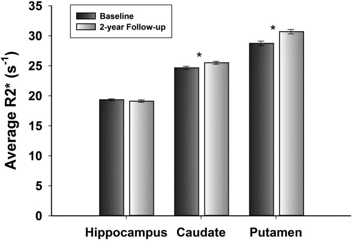
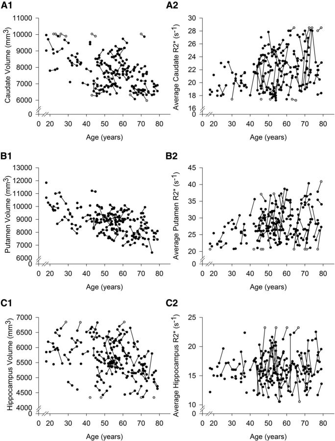
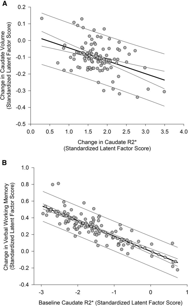
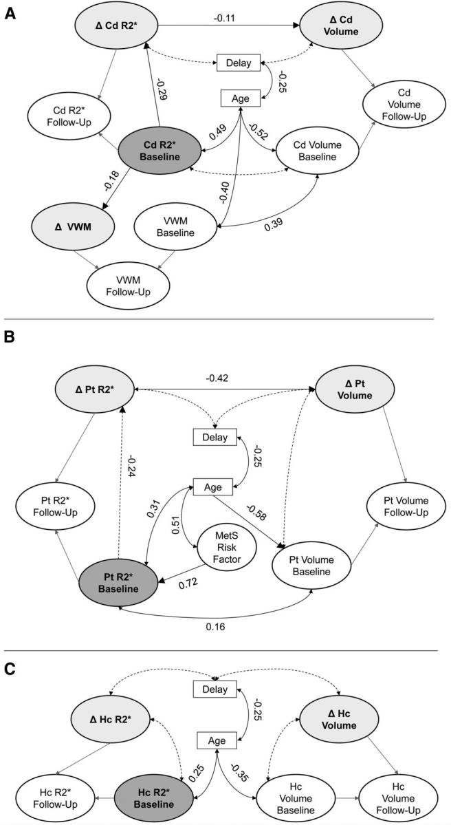
The accumulation of non-heme iron in the brain has been proposed as a harbinger of neural and cognitive decline in aging and neurodegenerative disease, but support for this proposal has been drawn from cross-sectional studies, which do not provide valid estimates of change. Here, we present longitudinal evidence of subcortical iron accumulation in healthy human adults (age 19-77 at baseline). We used R2* relaxometry to estimate regional iron content twice within a 2 year period, measured volumes of the striatum and the hippocampus by manual segmentation, and assessed cognitive performance by working memory tasks. Two-year change and individual differences in the change of regional volumes, regional iron content, and working memory were examined by latent change score models while taking into account the age at baseline and metabolic risk indicators. Over the examined period, volume reduction occurred in the caudate nucleus and hippocampus, but iron content increased only in the striatum, where it explained shrinkage. Higher iron content in the caudate nucleus at baseline predicted lesser improvement in working memory after repeat testing. Although advanced age and elevated metabolic syndrome risk were associated with greater iron content in the putamen at baseline, neither age nor metabolic risk influenced change in any variable. Thus, longitudinal evidence supports the notion that accumulation of subcortical iron is a risk factor for neural and cognitive decline in normal aging.

Keywords: MRI; R2*; aging; brain; longitudinal; metabolic syndrome.


Figures

Figure 1.
Figure 1.
A flow chart depicting recruitment, sample selection, and attrition. A, Recruitment. Persons who answered the ads were contacted by telephone, interviewed, and selected and received detailed health questionnaires. B, Selection. The number of participants who passed the initial health screening and subsequent screening for MRI eligibility were scanned on the 4T scanner, did not have incidental findings, and did not develop exclusionary conditions or acquire MRI contraindications in the course of the longitudinal study. C, Attrition. The numbers of participants who were unable to start baseline measurements despite full eligibility and those who had baseline measurements but did not return at the 2 year follow-up are shown. For details of exclusionary criteria and screening instruments, see Materials and Methods.
Figure 2.
Figure 2.
Examples of manual tracings for R2* relaxometry and volumetry. For R2*, each ROI was manually traced on an anatomical reference image (first echo) and copied to T2* maps. On the T2*-weighted images, voxels representing high magnetic susceptibility (i.e., iron) are visualized with dark intensity. R2* measures were calculated as the reciprocal of T2* as measured from the image intensity. Noise was removed from measurements by applying a threshold. For volumetry, each ROI was manually traced on T1-weighted MPRAGE images aligned to optimize visualization of each structure in the coronal plane. See Materials and Methods for details. Blue, Caudate nucleus; green, putamen; red, hippocampus.
Figure 3.
Figure 3.
Example of a latent change score model for change in regional volume (Vol). An identical model was used for regional R2* measures, and the same modeling approach was applied to working memory and metabolic syndrome risk constructs. The asterisk indicates a cross-time constraint, and coefficients set to 1 were used to identify the latent factors and to compute latent change. β, Mean effect at baseline; α, variance at baseline; δ, mean change; γ, variance in change; ε, covariance of baseline effect with change after 2 years.
Figure 4.
Figure 4.
Average regional R2* at baseline and at follow-up after a 2 year delay. Error bars represent SEM. The asterisk indicates significant within-region change, p < 0.02. At baseline and follow-up, all between-region comparisons were significant (p < 0.001).
Figure 5.
Figure 5.
Individual change trajectories in volume (adjusted for intracranial volume and summed across the hemispheres) and iron content (R2* averaged across the hemispheres) in the caudate, putamen, and hippocampus as a function of baseline age. Longer R2* is interpreted as greater iron content. Gray circles represent winsorized data points. A1, A2, In the caudate, regional mean change and individual differences in change in iron and volume were significant (p ≤ 0.02; α′ = 0.02). B1, B2, In the putamen, change in iron content was significant (p ≤ 0.02; α′ = 0.02), but not change in volume; however, there were significant individual differences in both measures (p ≤ 0.02; α′ = 0.02). C1, C2, In the hippocampus, iron content did not change, and the change in volume was nominally significant (p = 0.03; α′ = 0.02), but individual differences in longitudinal stability of both measures were significant (p ≤ 0.02; α′ = 0.02).
Figure 6.
Figure 6.
The associations between iron content and volume change in the caudate nucleus and changes in verbal working memory. Standardized latent factor scores for each latent variable were extracted from the final model and, therefore, reflect measures that are free of error and residualized on covariates (baseline age, interval between measurement occasions, and control between baseline measures and longitudinal change). Prediction limits and 95% confidence limits are shown for the regression lines. A, Longitudinal increase in iron content is associated with decrease in volume after 2 years. Positive values of change in iron content correspond to an increase over time, and negative values of change in volume indicate shrinkage. B, Greater iron content in the caudate nucleus at baseline predicted lesser repeated-testing gains in verbal working memory after 2 years. Baseline measurements were normed before model estimation, and increasing values of latent baseline iron correspond to greater iron content; positive values of latent change in verbal working memory indicate improvement in performance after 2 years.
Figure 7.
Figure 7.
Final parallel process latent change score models of the effects in each region: A, caudate nucleus; B, putamen; C, hippocampus. Only the latent models are shown, and all specified effects are significant; the measurement models are not shown, and cross-time correlations between measurements were estimated as covariates that are not shown here. Latent change scores are shaded light gray, and the baseline iron factor is dark gray. Dashed lines indicate nonsignificant pathways that were maintained for covariate control. A, Greater baseline iron content in the caudate is indirectly associated with shrinkage (indirect effect, 0.03; p = 0.02) and predicts lesser repeated-testing gains in verbal working memory after 2 years. B, Greater baseline iron in the putamen was indirectly associated with shrinkage after 2 years (indirect effect, 0.10; p = 0.04), and greater baseline iron content is associated with increased MetS risk. C, Iron is not related to volume measures in the hippocampus. Δ, latent change; VWM, verbal working memory.

References

    1. Apter NS, Halstead WC, Heimburger RF. Impaired cerebral functions in essential hypertension. Am J Psychiatry. 1951;107:808–813. - PubMed
    1. Bäckman L, Nyberg L, Lindenberger U, Li SC, Farde L. The correlative triad among aging, dopamine, and cognition: current status and future prospects. Neurosci Biobehav Rev. 2006;30:791–807. - PubMed
    1. Bartzokis G. Alzheimer's disease as homeostatic responses to age-related myelin breakdown. Neurobiol Aging. 2011;32:1341–1371. doi: 10.1016/j.neurobiolaging.2009.08.007. - DOI - PMC - PubMed
    1. Bartzokis G, Lu PH, Tingus K, Peters DG, Amar CP, Tishler TA, Finn JP, Villablanca P, Altshuler LL, Mintz J, Neely E, Connor JR. Gender and iron genes may modify associations between brain iron and memory in healthy aging. Neuropsychopharmacology. 2011;36:1375–1384. doi: 10.1038/npp.2011.22. - DOI - PMC - PubMed
    1. Berg D, Kruger R, Rieß R, Riederer P. Parkinson's disease. In: Youdim M, Riederer P, Mandel S, Battistin L, editors. Handbook of neurochemistry and molecular neurobiology: degenerative diseases of the nervous system. Ed 3. New York: Springer; 2007. pp. 1–20.

Publication types