. 2015 Jul;89(14):7089-96.
doi: 10.1128/JVI.00821-15. Epub 2015 Apr 29.

The Nectin-4/Afadin Protein Complex and Intercellular Membrane Pores Contribute to Rapid Spread of Measles Virus in Primary Human Airway Epithelia

Affiliations

The Nectin-4/Afadin Protein Complex and Intercellular Membrane Pores Contribute to Rapid Spread of Measles Virus in Primary Human Airway Epithelia

Brajesh K Singh et al. J Virol. 2015 Jul.

Erratum in

Abstract

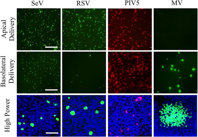
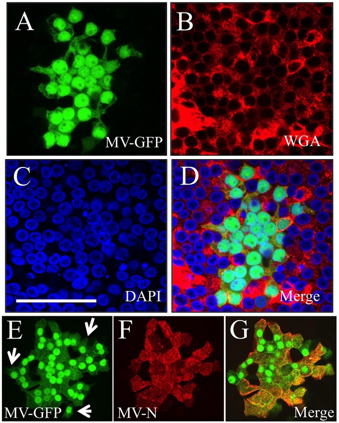
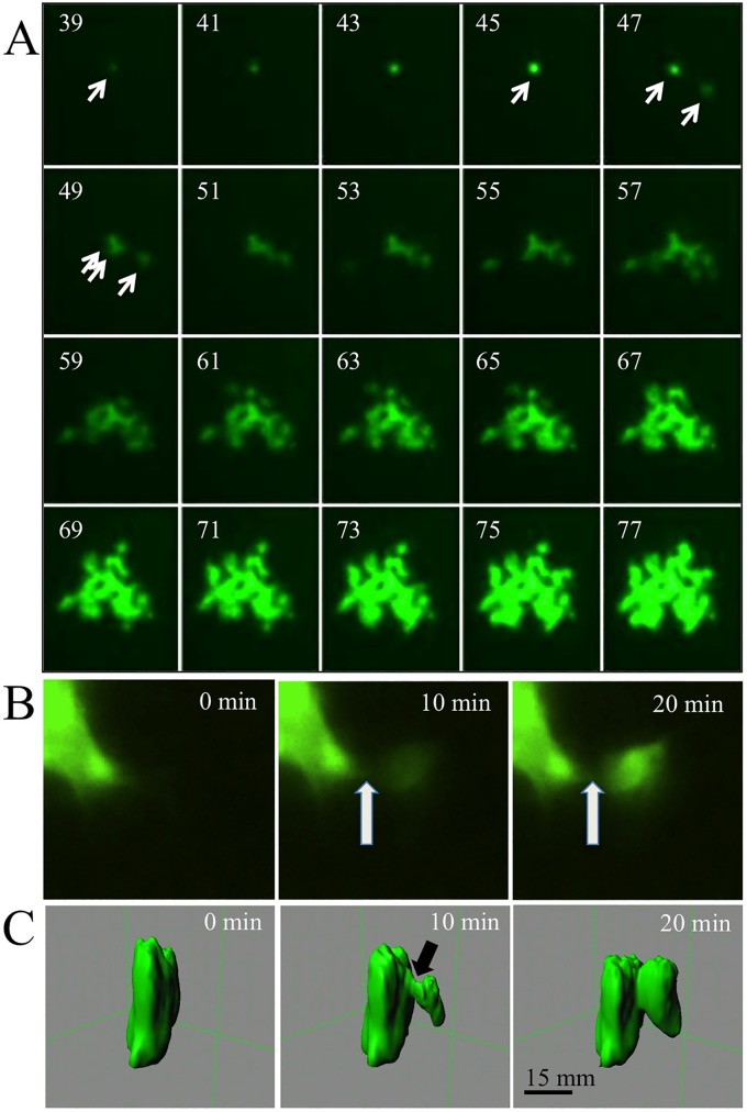
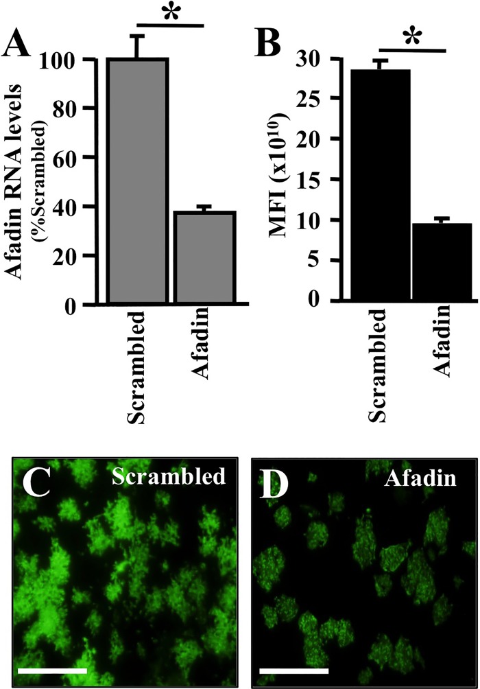
The discovery that measles virus (MV) uses the adherens junction protein nectin-4 as its epithelial receptor provides a new vantage point from which to characterize its rapid spread in the airway epithelium. We show here that in well-differentiated primary cultures of airway epithelial cells from human donors (HAE), MV infectious centers form rapidly and become larger than those of other respiratory pathogens: human respiratory syncytial virus, parainfluenza virus 5, and Sendai virus. While visible syncytia do not form after MV infection of HAE, the cytoplasm of an infected cell suddenly flows into an adjacent cell, as visualized through wild-type MV-expressed cytoplasmic green fluorescent protein (GFP). High-resolution video microscopy documents that GFP flows through openings that form on the lateral surfaces between columnar epithelial cells. To assess the relevance of the protein afadin, which connects nectin-4 to the actin cytoskeleton, we knocked down its mRNA. This resulted in more-limited infectious-center formation. We also generated a nectin-4 mutant without the afadin-binding site in its cytoplasmic tail. This mutant was less effective than wild-type human nectin-4 at promoting MV infection in primary cultures of porcine airway epithelia. Thus, in airway epithelial cells, MV spread requires the nectin-4/afadin complex and is based on cytoplasm transfer between columnar cells. Since the viral membrane fusion apparatus may open the passages that allow cytoplasm transfer, we refer to them as intercellular membrane pores. Virus-induced intercellular pores may contribute to extremely efficient measles contagion by promoting the rapid spread of the virus through the upper respiratory epithelium.

Importance: Measles virus (MV), while targeted for eradication, still causes about 120,000 deaths per year worldwide. The recent reemergence of measles in insufficiently vaccinated populations in Europe and North America reminds us that measles is extremely contagious, but the processes favoring its spread in the respiratory epithelium remain poorly defined. Here we characterize wild-type MV spread in well-differentiated primary cultures of human airway epithelial cells. We observed that viral infection promotes the flow of cytoplasmic contents from infected to proximal uninfected columnar epithelial cells. Cytoplasm flows through openings that form on the lateral surfaces. Infectious-center growth is facilitated by afadin, a protein connecting the adherens junction and the actin cytoskeleton. The viral fusion apparatus may open intercellular pores, and the cytoskeleton may stabilize them. Rapid homogenization of cytoplasmic contents in epithelial infectious centers may favor rapid spread and contribute to the extremely contagious nature of measles.


Figures

FIG 1
FIG 1
Paramyxovirus infection of HAE. (Top and center) Recombinant Sendai virus (SeV) expressing GFP, respiratory syncytial virus (RSV) expressing GFP, parainfluenza virus 5 (PIV5) expressing mCherry, and measles virus (MV) expressing GFP were applied to the apical (top row) or basolateral (center row) surface of HAE for 2 h at a multiplicity of infection of 1. Three days later, low-power images of live cells were collected with an inverted fluorescence microscope. Bar, 200 μm. (Bottom) High-power images of fixed cells collected by confocal microscopy. Bar, 50 μm.
FIG 2
FIG 2
MV does not form syncytia in HAE. HAE were infected from the basolateral surface with MV-GFP. Three days later, GFP expression was documented (A), the cell surface was stained with wheat germ agglutinin (WGA) (B), and nuclei were stained with 4′,6-diamidino-2-phenylindole (DAPI) (C). A merged image is shown (D). MV N protein colocalizes with GFP in infectious centers. Three days postinfection with MV-GFP, HAE were stained with anti-N protein antibody. GFP expression (E) and N protein staining (F) were observed in almost every MV-infected cell with few exceptions (white arrow). Panels E and F each represent a digitally compressed z-stack; panel G is a merged image of a single z-slice from panels E and F. Bars, 50 μm.
FIG 3
FIG 3
Time lapse microscopy analysis of MV infectious-center formation in HAE. HAE were infected from the basolateral surface with MV-GFP. (A) Growth of one infectious center shown at 2-h intervals starting at 39 h postinfection. An arrow in the 39-h panel indicates a single infected cell that infects another cell (at 47 h) and then a third cell (at 49 h). During the entire observation period, there was no visible cytopathic effect, and syncytium formation did not occur. (B and C) Higher-magnification, 10-min interval analyses of intercellular cytoplasmic GFP transfer at ∼48 h postinfection. (B) An inverted fluorescence microscope was used. (C) 3D reconstruction by confocal microscopy. The arrow indicates the point of initial GFP leakage.
FIG 4
FIG 4
Localization of AJ proteins in HAE. Fixed and permeabilized HAE were immunostained for the TJ marker ZO-1 (red) or afadin (green) (A) and for afadin (red) or nectin-4 (green) (B). (Top) xy en face views; (bottom) xz vertical views. Red and green arrows indicate the localizations of ZO-1 and afadin, respectively. White arrows indicate the colocalization of afadin and nectin-4, and yellow arrows indicate nectin-4 expression. Cell nuclei were visualized using DAPI (blue). Images were acquired with a confocal laser scanning microscope (Zeiss LSM 510).
FIG 5
FIG 5
Afadin knockdown reduces the efficiency of MV infection in HAE. Afadin was silenced in primary human airway epithelia by using a reverse transfection technique (35). (A) Afadin mRNA abundance following transfection with scrambled siRNA or afadin siRNA, as determined by RT-qPCR 24 h posttransfection. Twenty-four hours after siRNA transfection, MV-GFP was delivered to the basolateral surfaces of epithelial cells. Results are means for 3 experiments, each with samples from 3 donors; *, P < 0.001. (B) The mean fluorescence intensity (MFI) was measured using bioluminescence imaging 3 days after infection. (C and D) Representative images of MV spread, as monitored by GFP expression, in epithelia transfected with scrambled or afadin siRNA. Bars, 100 μm.
FIG 6
FIG 6
Relevance of the afadin-binding sequence in the nectin-4 cytoplasmic tail for MV epithelial infection. (A to C) Localization of human nectin-4 in PAE either transduced from the basolateral surface with an adenoviral vector expressing human nectin-4 (A) or human N4ΔGHLV (B) or mock transduced (C). Two days later, cells were fixed with 2% paraformaldehyde, permeabilized with 0.2% Triton X-100, and incubated overnight with rabbit polyclonal antibodies against human nectin-4. Nectin-4 was visualized with a secondary antibody conjugated to Alexa Fluor 546 (red). Cell nuclei were visualized using DAPI (blue). For each panel, both an xy en face view (top) and an xz vertical view (bottom) are shown. Images were acquired with a confocal laser scanning microscope. White arrows indicate basolateral localization; yellow arrows indicate junctional expression. (D) Analysis of Ad-nectin-4 and Ad-N4ΔGHLV expression levels in CHO cells. β-tub., β-tubulin. (E) PAE transduced with an adenoviral vector expressing either human nectin-4 or N4ΔGHLV were infected from either the apical or the basolateral surface with MV-GFP, and the infectious centers were counted. An adenoviral vector expressing mCherry served as a negative control. Results are means for 3 experiments, each with samples from 3 donors; *, P < 0.01.

References

    1. Mateo M, Navaratnarajah CK, Cattaneo R. 2014. Structural basis of efficient contagion: measles variations on a theme by parainfluenza viruses. Curr Opin Virol 5:16–23. doi:10.1016/j.coviro.2014.01.004. - DOI - PMC - PubMed
    1. Chen SY, Anderson S, Kutty PK, Lugo F, McDonald M, Rota PA, Ortega-Sanchez IR, Komatsu K, Armstrong GL, Sunenshine R, Seward JF. 2011. Health care-associated measles outbreak in the United States after an importation: challenges and economic impact. J Infect Dis 203:1517–1525. doi:10.1093/infdis/jir115. - DOI - PubMed
    1. Tatsuo H, Ono N, Tanaka K, Yanagi Y. 2000. SLAM (CDw150) is a cellular receptor for measles virus. Nature 406:893–897. doi:10.1038/35022579. - DOI - PubMed
    1. Ferreira CS, Frenzke M, Leonard VH, Welstead GG, Richardson CD, Cattaneo R. 2010. Measles virus infection of alveolar macrophages and dendritic cells precedes spread to lymphatic organs in transgenic mice expressing human signaling lymphocytic activation molecule (SLAM, CD150). J Virol 84:3033–3042. doi:10.1128/JVI.01559-09. - DOI - PMC - PubMed
    1. Lemon K, de Vries RD, Mesman AW, McQuaid S, van Amerongen G, Yuksel S, Ludlow M, Rennick LJ, Kuiken T, Rima BK, Geijtenbeek TB, Osterhaus AD, Duprex WP, de Swart RL. 2011. Early target cells of measles virus after aerosol infection of non-human primates. PLoS Pathog 7:e1001263. doi:10.1371/journal.ppat.1001263. - DOI - PMC - PubMed

Publication types

MeSH terms

LinkOut - more resources