Review
. 2015 Jun;79(2):225-41.
doi: 10.1128/MMBR.00061-14.

Activation and regulation of DNA-driven immune responses

Affiliations
Review

Activation and regulation of DNA-driven immune responses

Søren R Paludan. Microbiol Mol Biol Rev. 2015 Jun.

Abstract

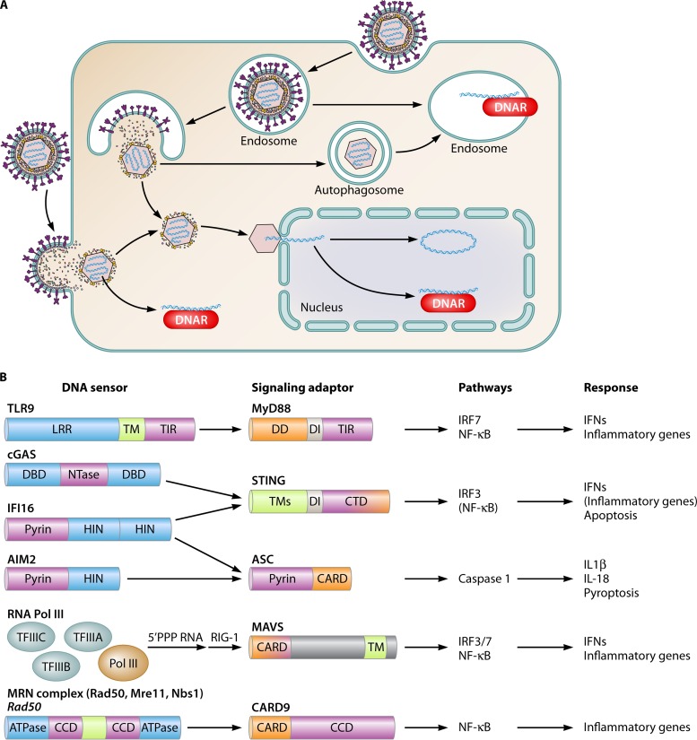
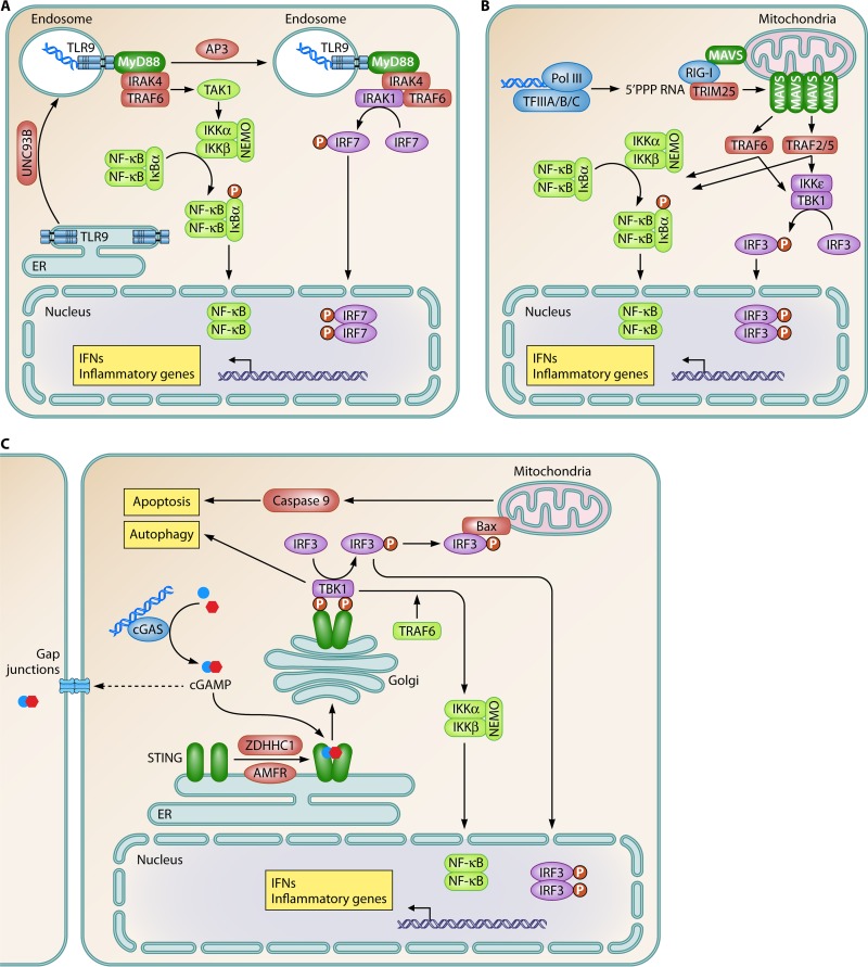
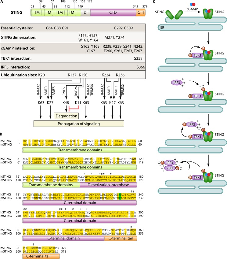
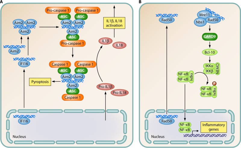
The innate immune system provides early defense against infections and also plays a key role in monitoring alterations of homeostasis in the body. DNA is highly immunostimulatory, and recent advances in this field have led to the identification of the innate immune sensors responsible for the recognition of DNA as well as the downstream pathways that are activated. Moreover, information on how cells regulate DNA-driven immune responses to avoid excessive inflammation is now emerging. Finally, several reports have demonstrated how defects in DNA sensing, signaling, and regulation are associated with susceptibility to infections or inflammatory diseases in humans and model organisms. In this review, the current literature on DNA-stimulated innate immune activation is discussed, and important new questions facing this field are proposed.


Figures

FIG 1
FIG 1
Innate DNA sensors and subcellular localization of DNA recognition. (A) DNA sensors are expressed in endosomes, the cytoplasm, and the nucleus. DNA may come into contact with DNA sensors in the endosome through uptake via endosomal or autophagy pathways. Stimulation of the cytoplasmic DNA sensing pathways may occur as a result of (i) bacteriolysis or viral capsid degradation in the cytosol, (ii) accumulation of endogenous retroviral reverse transcripts or DNA replication intermediates, or (iii) leakage of DNA from endosomal compartments. In the nucleus, the features that distinguish stimulatory from nonstimulatory DNAs are not known. Factors that may be involved include epigenetic modifications and interactions with histones. DNAR, DNA receptor. (B) DNA sensors mediate signaling via adaptor proteins to activate specific pathways and biological responses. Blue, DNA-binding domains; purple, domains/proteins relaying signals between the sensors and the adaptor proteins; orange, domains relaying signals from the adaptor proteins and downstream. CARD, caspase activation and recruitment domain; CCD, coiled-coil domain; CTD, C-terminal domain; DD, death domain; DI, dimerization interphase; LLR, leucine-rich repeat; TIR, Toll/interleukin-1 receptor domain; TM, transmembrane region.
FIG 2
FIG 2
Signal transduction pathways activated by DNA to stimulate type I IFN expression. Innate DNA sensing induces the activation of three signaling pathways to stimulate type I IFN expression. (A) TLR9; (B) RNA polymerase III; (C) cGAS. ER, endoplasmic reticulum. Blue, sensor; green, adaptor; purple, IRF pathway; light green, NF-κB pathway; claret, other signaling molecules; yellow boxes, biological effector functions.
FIG 3
FIG 3
STING, the adaptor protein in DNA-activated signaling for IFN expression. (A) Summary of the known molecular details on the residues and regions of STING involved in the biological functions of the molecule. CTT, C-terminal tail. (B) Alignment of human STING (hSTING) and murine STING (mSTING), which have 63% sequence homology. Position 232 (position 231 in murine STING), which contributes critically to the differential responsiveness of human and murine STINGs to 3′,5′-linked CDNs, is highlighted in green. Conserved residues are highlighted in yellow. Residues involved in STING dimerization are marked with *. Residues involved in cGAMP binding are marked with #. Serine residues in the C-terminal tail involved in the recruitment of TBK1 and IRF3 are shown in boldface type and marked with ¤ and §, respectively. (C) Model for IRF3 activation through the cGAS-STING pathway. In the first step, cGAMP binding induces a conformational change in the C-terminal domain of STING. The second step involves the recruitment and activation of TBK1, followed by TBK1-mediated phosphorylation of STING at serine 366. The third step is the recruitment of monomeric IRF3 to STING at phosphoserine 366, through an interaction with a positively charged surface on IRF3. The fourth step is TBK1-mediated phosphorylation of IRF3 at C-terminal serines, including positions 385 and 386. The fifth step involves the homodimerization of phosphorylated IRF3 molecules and the activation of transcriptional functions.
FIG 4
FIG 4
Signal transduction pathways activated by DNA to stimulate activation of inflammasomes and the NF-κB pathway. (A) AIM2-like receptors; (B) Rad50. Blue, sensor; green, adaptor; light green, NF-κB pathway; orange, inflammasome pathway; claret, other signaling molecules; yellow boxes, biological effector functions.
FIG 5
FIG 5
Endogenous and microbial mechanisms to downmodulate and evade DNA-driven innate immune responses. (A) DNA is degraded by DNase II in lysosomal compartments and by Trex1 in the cytoplasm. DNA is also targeted for degradation by an autophagy pathway stimulated upon DNA binding by cGAS, leading to the release of Rubicon from Beclin-1. In addition to DNA turnover, the pathway is also regulated at the level of STING. After activation, this protein can be degraded through either proteasomal or autophagocytic pathways. Moreover, caspases 3, 7, and 9, activated during mitochondrial apoptosis, suppress DNA-stimulated IFN production by targeting a step upstream of TBK1 phosphorylation. Finally, the ULK1 kinase is activated by DNA and cGAMP to catalyze specific serine phosphorylation of STING in the C-terminal domain, leading to the downmodulation of the IRF3 pathway. (B) The SKIV2L exosome pathway degrades RNA that stimulates the RIG-I pathway and is thus likely to also be involved in the control of the Pol III–RIG-I pathway. (C) Viruses evade and counteract DNA-driven immune responses at several levels, including prevention of the release of DNA into the cytoplasm, degradation of specific DNA sensors, and inhibition of signaling proteins via protein-protein interactions.

References

    1. Medzhitov R. 2008. Origin and physiological roles of inflammation. Nature 454:428–435. doi: 10.1038/nature07201. - DOI - PubMed
    1. Rotem Z, Cox RA, Isaacs A. 1963. Inhibition of virus multiplication by foreign nucleic acid. Nature 197:564–566. doi: 10.1038/197564a0. - DOI - PubMed
    1. Hemmi H, Takeuchi O, Kawai T, Kaisho T, Sato S, Sanjo H, Matsumoto M, Hoshino K, Wagner H, Takeda K, Akira S. 2000. A Toll-like receptor recognizes bacterial DNA. Nature 408:740–745. doi: 10.1038/35047123. - DOI - PubMed
    1. Ishii KJ, Coban C, Kato H, Takahashi K, Torii Y, Takeshita F, Ludwig H, Sutter G, Suzuki K, Hemmi H, Sato S, Yamamoto M, Uematsu S, Kawai T, Takeuchi O, Akira S. 2006. A Toll-like receptor-independent antiviral response induced by double-stranded B-form DNA. Nat Immunol 7:40–48. doi: 10.1038/ni1282. - DOI - PubMed
    1. Stetson DB, Medzhitov R. 2006. Recognition of cytosolic DNA activates an IRF3-dependent innate immune response. Immunity 24:93–103. doi: 10.1016/j.immuni.2005.12.003. - DOI - PubMed

Publication types

LinkOut - more resources