. 2015 Apr 14:6:158.
doi: 10.3389/fimmu.2015.00158. eCollection 2015.

Negative Regulation of Humoral Immunity Due to Interplay between the SLAMF1, SLAMF5, and SLAMF6 Receptors

Affiliations

Negative Regulation of Humoral Immunity Due to Interplay between the SLAMF1, SLAMF5, and SLAMF6 Receptors

Ninghai Wang et al. Front Immunol. .

Abstract

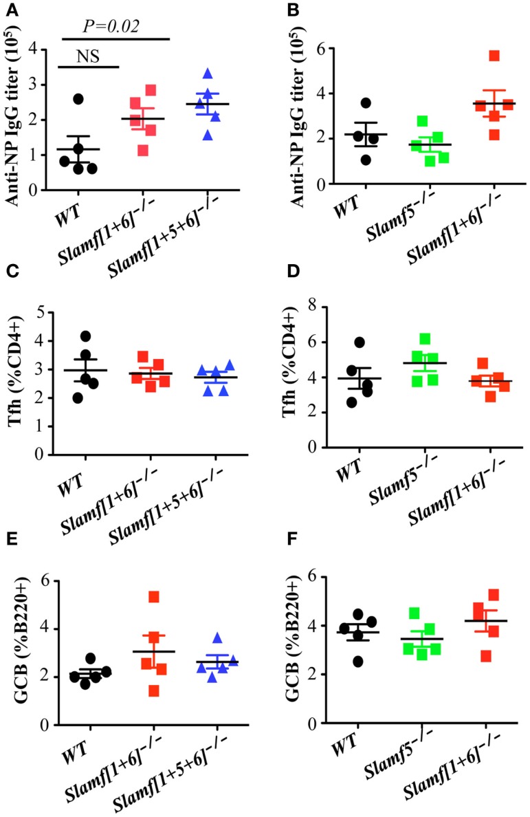
Whereas the SLAMF-associated protein (SAP) is involved in differentiation of T follicular helper (Tfh) cells and antibody responses, the precise requirements of SLAMF receptors in humoral immune responses are incompletely understood. By analyzing mice with targeted disruptions of the Slamf1, Slamf5, and Slamf6 genes, we found that both T-dependent and T-independent antibody responses were twofold higher compared to those in single knockout mice. These data suggest a suppressive synergy of SLAMF1, SLAMF5, and SLAMF6 in humoral immunity, which contrasts the decreased antibody responses resulting from a defective GC reaction in the absence of the adapter SAP. In adoptive co-transfer assays, both [Slamf1 + 5 + 6] (-/-) B and T cells were capable of inducing enhanced antibody responses, but more pronounced enhancement was observed after adoptive transfer of [Slamf1 + 5 + 6] (-/-) B cells compared to that of [Slamf1 + 5 + 6] (-/-) T cells. In support of [Slamf1 + 5 + 6] (-/-) B cell intrinsic activity, [Slamf1 + 5 + 6] (-/-) mice also mounted significantly higher antibody responses to T-independent type 2 antigen. Furthermore, treatment of mice with anti-SLAMF6 monoclonal antibody results in severe inhibition of the development of Tfh cells and GC B cells, confirming a suppressive effect of SLAMF6. Taken together, these results establish SLAMF1, SLAMF5, and SLAMF6 as important negative regulators of humoral immune response, consistent with the notion that SLAM family receptors have dual functions in immune responses.

Keywords: SLAM family receptors; SLAM-associated protein; anti-SLAMF6 mAb; follicular T helper cells; germinal center B cells; marginal zone B cells.


Figures

Figure 1
Figure 1
Generation of Slamf[1 + 6]−/− and Slamf[1 + 5 + 6]−/− mice. (A) Schematic representation of the Slamf[1 + 6] and Slamf[1 + 5 + 6] targeting strategy. Top: illustration of the genomic mouse SLAMF1-5-6 locus after targeted replacement of exon 2 and 3 of both Slamf1 and Slamf6 genes. Middle: The Slamf1-5-6 locus after Cre-mediated recombination leading to the deletion of the LoxP site-flanked genomic fragment. Bottom: PCR genotyping primers (PF and PR) used to confirm the junction of Cre-mediated deletion from mouse-tail DNA. (B) Thymocytes from WT, Slamf[1 + 5]/, and Slamf[1 + 5 + 6]/ mice were stained with anti-SLAMF1, anti-SLAMF5, and anti-SLAMF6 antibodies, and the expression of SLAMF1, SLAMF5, and SLAMF6 was evaluated by flow cytometry.
Figure 2
Figure 2
Comparison of B cell and T cell populations in Slamf[1 + 5]−/−, Slamf[1 + 5 + 6]−/−, and WT mice. Flow cytometric analyses of B cell and T cell subsets in the spleens of Slamf[1 + 6]/, Slamf[1 + 5 + 6]/, and WT mice: (A) CD19+AA4-IgMhi cells in spleens are gated for the expression of CD23 and CD21 to delineate CD21+CD23- marginal zone (MZ) B cells. (B) Percentage of CD19+AA4 IgMhiCD21+ CD23 MZ B cells. (C) The number of CD19+AA4 IgMhiCD21+CD23- MZ B cells. (D) Splenocytes from Slamf[1 + 6]/, Slamf[1 + 5 + 6]/, and WT mice are stained for surface expression of CD3 and CD19. (E) CD19+ AA4 IgMhiCD21 cells in spleens are gated for the expression of CD23 and CD21 to delineate CD21+CD23+ follicular B cells in the spleens. (F) Transitional B cell subsets in spleens are stained for the expression of CD19, AA4.1, CD23, and IgM: T1 (CD19+AA4+IgMhiCD23-), T2 (CD19+AA4+IgMhiCD23+), and T3 (CD19+AA4+IgMloCD23-).
Figure 3
Figure 3
A combination of SLAMF1, SLAMF5, and SLAMF6 negatively regulates T cell dependent antibody responses, but normal Tfh and GCB development is observed in Slamf[1 + 5 + 6]−/− mice. WT, Slamf5/, Slamf[1 + 6]/, and Slamf[1 + 5 + 6]/ mice were immunized with 40 μg of NP-OVA and serum was collected on day 9. (A) NP-specific IgG titers for Slamf[1 + 6]/, Slamf[1 + 5 + 6]/, and WT mice immunized with NP-OVA in CFA were determined by ELISA using NP(4)-BSA coated plates. (B) NP-specific IgG titers for Slamf5/, Slamf[1 + 6]/, and WT mice immunized with NP-OVA in Alum were determined by ELISA using NP(4)-BSA coated plates. (C) Percentage of Tfh cells (CD4+PD-1+CXCR5+) in the spleens of Slamf[1 + 6]/, Slamf[1 + 5 + 6]/, and WT mice. (D) Percentage of Tfh cells (CD4+PD-1+CXCR5+) in the spleens of Slamf5/, Slamf[1 + 6]/, and WT mice. (E) Percentage of Germinal Center B cells (B220+GL7+Fas+) in the spleens of Slamf[1 + 6]/, Slamf[1 + 5 + 6]/, and WT mice. (F) Percentage of Germinal Center B cells (B220+GL7+Fas+) in the spleens of Slamf5/, Slamf[1 + 6]/, and WT mice. Data represent at least three independent experiments.
Figure 4
Figure 4
A combined absence of SLAMF1, SLAMF5, and SLAMF6 enhances antigen specific plasma cell expansion. Slamf[1 + 6]/, Slamf[1 + 5 + 6]/, and WT mice were immunized with NP-OVA in CFA. After 9 days, spleens were isolated and subjected to staining with the indicated antibodies and analyzed by flow cytometry. (A) Representative FACS plots showing B220+IgD-CD138+ plasma cells from the spleens of Slamf[1 + 6]/, Slamf[1 + 5 + 6]/, and WT mice. (B) Percentage of Plasma cells (B220+IgD-CD138+) from the spleens of Slamf[1 + 6]/, Slamf[1 + 5 + 6]/, and WT mice. (C) Representative FACS plots showing B220+NP+IgD- NP-specific plasma cells from the spleens of Slamf[1 + 5 + 6]/ and WT mice. (D) Percentage of NP-staining plasma cells (B220+NP+IgD-) from the spleens of Slamf[1 + 5 + 6]/ and WT mice. Data represent at least three independent experiments.
Figure 5
Figure 5
The adoptive transfer of naïve Slamf[1 + 5 + 6]−/− T or B cells enhanced NP-specific antibody responses after co-transfer of WT B or T cells into Rag-1−/− mice. CD4+ T cells (5 × 106) together with 10 × 106 B cells are isolated from WT and Slamf[1 + 5 + 6]−/− mice and transferred into Rag-1−/− recipients in the following four combinations of T and B cells: WT CD4+ T and WT B cells, Slamf[1 + 5 + 6]−/− CD4+ T and Slamf[1 + 5 + 6]−/− B cells, WT CD4+ T and Slamf[1 + 5 + 6]−/− B cells, and Slamf[1 + 5 + 6]−/− CD4+ T and WT B cells. The Rag-1−/− recipients were immunized with 40 μg of NP-OVA in CFA 7 days after the transfer. Mice were sacrificed and NP-specific IgG titers were determined by ELISA day 9 post-immunization. Data are representative of three independent experiments.
Figure 6
Figure 6
A combined ablation of SLAMF1, SLAMF5, and SLAMF6 shows a selective increase in MZ B cells and enhanced TI-2 antibody responses. WT, Slamf[1 + 6]−/−, and Slamf[1 + 5 + 6]−/− mice were immunized with 20 μg of NP-Ficoll. NP-specific IgM (A) and IgG3 (B) titers were determined at day 7 by ELISA after serial dilutions of the serum. Results are representative of three independent experiments.
Figure 7
Figure 7
Administration of anti-SLAMF6 antibody has a negative effect on antibody production in protein-immunized WT mice. Mice were immunized with 40 μg NP-OVA in CFA and some were injected with either 250 μg of anti-SLAMF6 (330), anti-SLAMF1 (9D1), anti-SLAMF5 (M5), or mouse Ig isotype control. The mice were sacrificed on day 9 and serum was collected to measure Ig production. (A) NP-specific IgG titers from sera of anti-SLAMF6 injected mice were determined by ELISA. (B) Affinity of NP-specific IgG in immune-sera collected as in (A). (C) NP-specific IgM titers from sera of anti-SLAMF6 injected mice were determined by ELISA. (D) Affinity of NP-specific IgM in immune-sera collected as in (C). (E) NP-specific IgG titers from sera of anti-SLAMF1 injected mice were determined by ELISA. (F) NP-specific IgG titers from sera of anti-SLAMF5 injected or non-injected immunized mice were determined by ELISA. Results are representative of three independent experiments.
Figure 8
Figure 8
Administration of anti-SLAMF6 (330) antibody has a negative effect on GC B cell differentiation in protein-immunized WT mice. Mice were immunized with 40 μg of NP-OVA in CFA and some mice were injected with either 250 μg anti-SLAMF6 (330) or Ig isotype control. The mice were sacrificed on day 9. (A) Representative flow cytometry staining of B220+GL7+Fas+ Germinal Center B cells in the spleens of anti-SLAMF6, isotype, and non-injected immunized mice. (B) Percentage of Germinal Center B cells (B220+GL7+Fas+) in the spleens of anti-SLAMF6, isotype, and non-injected immunized mice was determined by flow cytometry. (C) The numbers of Germinal Center B cells (B220+GL7+Fas+) in the spleens of anti-SLAMF6, isotype, and non-injected immunized mice were determined by flow cytometry. (D) Representative of flow cytometry staining of B220+IgD-CD138+ plasma cells in the spleens of anti-SLAMF6, isotype, and non-injected immunized mice. (E) Percentage of plasma cells (B220+IgD-CD138+) in the spleens of anti-SLAMF6, isotype, and non-injected immunized mice. (F) Percentage of B220+CD86+ activated B cells in the spleens of anti-SLAMF6, isotype, and non-injected immunized mice. Results are representative of three independent experiments.
Figure 9
Figure 9
Administration of anti-SLAMF6 (330) antibody has a negative effect on Tfh cell differentiation in protein-immunized WT mice. Mice were immunized with 40 μg of NP-OVA in CFA and some mice were injected with either 250 μg anti-SLAMF6 (330) or Ig isotype control. The mice were sacrificed on day 9. (A) Representative flow cytometry staining of CD4+PD-1+CXCR5+ Tfh cells in the spleens of anti-SLAMF6, isotype, and non-injected immunized mice. (B) Percentage of Tfh cells (CD4+PD-1+CXCR5+) was determined by flow cytometry in the spleens of anti-SLAMF6, isotype, and non-injected immunized mice. (C) The number of Tfh cells (CD4+PD-1+CXCR5+) was determined by flow cytometry in the spleens of anti-SLAMF6, isotype, and non-injected immunized mice. (D) Percentage of CD4+CD44hiCD62lo memory T cells in the spleens of anti-SLAMF6, isotype, and non-injected immunized mice. (E) Percentage of CD4+CD44hiCD69hi activated T cells in the spleens of anti-SLAMF6, isotype, and non-injected immunized mice. Results are representative of three independent experiments.
Figure 10
Figure 10
Administration of anti-SLAMF6 (330) F(ab’)2 antibody has a similar negative effect on GC B cell and Tfh cell development in protein-immunized WT mice. Mice were immunized with 40 μg of NP-OVA in CFA and some were injected with 250 μg of anti-SLAMF6 F(ab’)2 on day 0 and day 4. The mice were sacrificed on day 9 and serum was collected to measure IgG production. (A) NP-specific IgG titers were determined by ELISA. (B–C) The percentage and number of Germinal Center B cells (B220+GL7+Fas+) were determined by flow cytometry. (D–E) The percentage and number of Tfh cells (CD4+PD-1+CXCR5+) were determined by flow cytometry. Results are representative of three independent experiments.
Figure 11
Figure 11
A model of SLAMF receptors: SHP1/2 action on the BCR during B cell activation. When B cells are activated, the ITSMs of SLAMF1, SLAMF5, and SLAMF6 recruit SHP1/2 and translocate these phosphatases to the vicinity of the B cell antigen receptor. Signaling from the BCR is thus down-regulated, maintaining proper response to antigens in humoral responses. When SLAMF1, SLAMF5, and SLAMF6 are deleted from B cells, inhibitory signaling mediated by SLAMF and SHP1/2 is dampened, which induces enhanced humoral responses.

References

    1. Rajewsky K. Clonal selection and learning in the antibody system. Nature (1996) 381:751–810.1038/381751a0 - DOI - PubMed
    1. Victora GD, Schwickert TA, Fooksman DR, Kamphorst AO, Meyer-Hermann M, Dustin ML, et al. Germinal center dynamics revealed by multiphoton microscopy with a photoactivatable fluorescent reporter. Cell (2010) 143:592–605.10.1016/j.cell.2010.10.032 - DOI - PMC - PubMed
    1. Vinuesa CG, Linterman MA, Goodnow CC, Randall KL. T cells and follicular dendritic cells in germinal center B-cell formation and selection. Immunol Rev (2010) 237:72–89.10.1111/j.1600-065X.2010.00937.x - DOI - PubMed
    1. Crotty S. Follicular helper CD4 T cells (TFH). Annu Rev Immunol (2011) 29:621–63.10.1146/annurev-immunol-031210-101400 - DOI - PubMed
    1. Victora GD, Nussenzweig MC. Germinal centers. Annu Rev Immunol (2012) 30:429–5710.1146/annurev-immunol-020711-075032 - DOI - PubMed