Case Reports
. 2015 Jan 31;9(1):36-42.
doi: 10.3941/jrcr.v9i1.2066. eCollection 2015 Jan.

Inflammatory pseudotumor of the urinary bladder

Affiliations
Case Reports

Inflammatory pseudotumor of the urinary bladder

Elsa Rosado et al. J Radiol Case Rep. .

Abstract

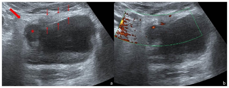
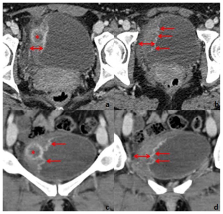
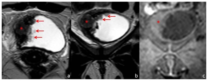
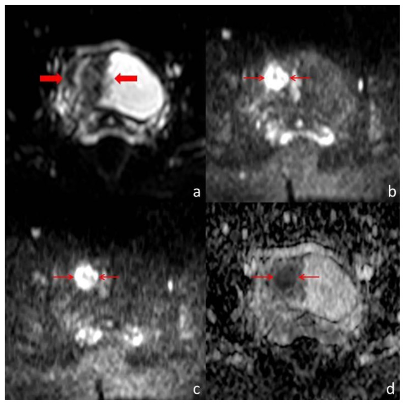
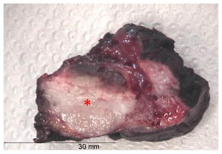
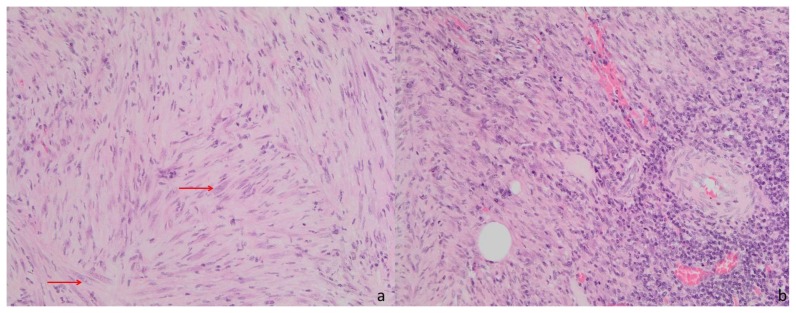
We report a case of an inflammatory pseudotumor of the urinary bladder in a 31 year-old woman. She presented at the emergency room with low abdominal pain and urinary symptoms. Abdominal ultrasound, computed tomography and magnetic resonance imaging were performed and revealed asymmetric thickening of the urinary bladder wall. Cystoscopy with urinary cytology revealed a benign nature of the process. The patient underwent partial cystectomy and the pathologic examination of the specimen revealed an inflammatory pseudotumor. We reviewed the clinical, imaging and pathological features of the inflammatory pseudotumor of the urinary bladder and discussed its differential diagnosis.

Keywords: Inflammatory pseudotumor; computed tomography; inflammatory myofibroblastic tumor; magnetic resonance imaging; plasma cell granuloma; ultrasound; urinary bladder.


Figures

Figure 1
Figure 1
Thirty-one year old woman with inflammatory pseudotumor of the urinary bladder. Abdominal US scan performed with a convex array transducer (3–6 MHz) on a Toshiba Aplio 500®. Axial scanning plane through the hypogastrium (a) demonstrates the partially filled urinary bladder, with a thickened wall (thin arrows). Maximum thickness was 21mm. In the right-anterior segment of the thickened wall there is a round, hypoechoic area (*) of 19mm diameter. The surrounding fat is hyperechoic (thick arrow). Power Doppler imaging in the same axial scanning plane (b) shows vascularization within the thickened wall.
Figure 2
Figure 2
Thirty-one year old woman with inflammatory pseudotumor of the urinary bladder. Contrast-enhanced CT, performed with a SimensSensation® (64 slice-CT). Radiation dose: 120Kv and 70 mAs. Injection of 120ml of 400mg/ml iodine concentration non-ionic contrast agent (Iomeron®). Images selected were taken 60s after contrast injection. Axial 1,5 thickness images (a and b) and coronal 4mm thickness reconstruction images (c and d) demonstrate right lateral bladder wall thickening (double arrow). Maximum thickness is 21mm. The mucosal surface is rim enhancing and has multiple papillary projections protruding into the urinary bladder lumen. Within the thickened wall, there is a nodular, 20mm diameter hypoenhancing area (*) with hyperenhancing margins.
Figure 3
Figure 3
Thirty-one year old woman with inflammatory pseudotumor of the urinary bladder. Pelvic MR performed with a Philips Intera® (1,5 T). T1-weighted images were obtained by 3D spoiled turbo gradient echo technique (TR/TE: 3,25/1,55 ms) with SPAIR fat suppression and SENSE acceleration (factor 2). T2-weighted images were obtained by spin-echo technique (TR/TE: 8148/150 ms). Axial T2-weighted (a), coronal T2-weighted (b) and coronal T1-weighted (c) images demonstrate the right urinary bladder wall thickening with a nodular solid area (*) of intermediate signal on the T2-weighted images and which is almost imperceptible on the T1-weighted image. The mucosal surface of the thickened wall has multiple papillary projections, well depicted on the T2-weighted images (arrows).
Figure 4
Figure 4
Thirty-one year old woman with inflammatory pseudotumor of the urinary bladder. Pelvic MR performed with a Philips Intera® (1,5 T). Diffusion-weighted images were obtained by echo planar imaging technique, with a TR/TE/TI of 1300/180/90 ms and a b value of 0 s/mm2 (a), 1000 s/mm2 (b) and 1500 s/mm2 (c). ADC maps were built based on the three b values (d). The parietal thickening (thick arrows) is well depicted on (a). At higher b values, the diffusion restriction of the nodular component of the lesion is evident (thin arrows), demonstrating its high cellularity. This area has lower signal intensity in the ADC map, confirming restricted diffusion.
Figure 5
Figure 5
Thirty-one year old woman with inflammatory pseudotumor of the urinary bladder. Gross examination of the surgical specimen. The resected piece measured 4,5×4×2cm. The lesion had 2,3×2×2cm and it was completely resected. In cross section (*) it was white and firm.
Figure 6
Figure 6
Thirty-one year old woman with inflammatory pseudotumor of the urinary bladder. Pathological study of the surgical specimen (hematoxylin and eosin staining, magnification: 20×). Spindle cell proliferation (a) with sparse mild nuclear atypia (arrows). Predominantly mononuclear inflammatory infiltrate (b).

References

    1. Park SB, Cho KS, Kim JK, et al. Inflammatory pseudotumor (myoblastic tumor) of the genitourinary tract. AJR Am J Roentgenol. 2008;191(4):1255–62. - PubMed
    1. Kumar A, Bhatti SS, Sharma S, Gupta SD, Kumar R. Inflammatory pseudotumor of urinary bladder - a diagnostic and management dilemma. Int Urol Nephrol. 2007;39(3):799–802. - PubMed
    1. Iczkowski KA, Shanks JH, Gadaleanu V, et al. Inflammatory pseudotumor and sarcoma of urinary bladder: differential diagnosis and outcome in thirty-eight spindle cell neoplasms. Mod Pathol. 2001;14(10):1043–51. - PubMed
    1. Wong-You-Cheong JJ, Woodward PJ, Manning MA, Davis CJ. From the archives of the AFIP: Inflammatory and nonneoplastic bladder masses: radiologic-pathologic correlation. Radiographics. 2006;26(6):1847–68. - PubMed
    1. Sugita R, Saito M, Miura M, Yuda F. Inflammatory pseudotumour of the bladder: CT and MRI findings. Br J Radiol. 1999;72(860):809–11. - PubMed

Publication types