Review
. 2015 Spring;8(2):99-109.

Mucosal histopathology in celiac disease: a rebuttal of Oberhuber's sub-division of Marsh III

Affiliations
Review

Mucosal histopathology in celiac disease: a rebuttal of Oberhuber's sub-division of Marsh III

Michael N Marsh et al. Gastroenterol Hepatol Bed Bench. 2015 Spring.

Abstract

Individuals with particular genetic backgrounds develop immune responses to wheat proteins and become 'gluten-sensitised'. Mucosal pathology arises through activated mucosal T lymphocytes, resulting in a graded, adverse reaction between particular genes and wheat proteins. Given these varied influences, the Marsh Classification broadly itemises those stages through which a normal mucosa (Marsh 0) evolves in becoming 'flat' (Marsh I, II, III). Recently, Oberhuber and colleagues suggested that Marsh III lesions required subdividing into a, b, c categories. We critically examined these subdivisions by means of correlative light and scanning electron microscopy (SEM). Our results demonstrate that Oberhuber's classification is untenable. In our view deriving from our observations, the artificial subdivisions proposed by those authors actually reflect misinterpretations of the true architectural contours of flat mucosae. Although these workers refer to "villous projections", SEM demonstrates that no such structures are present on flat - or immediately recovering - mucosae. Our data revealed on the surfaces of flat (Marsh III) mucosae, large open "basins", surrounded by raised collars - the latter, when viewed in histological section, being easily misconstrued as "villi". It seems that with subsequent upward growth, these collars coalesce into low ridges, thence becoming broader and higher convolutions. It is noticeable that there are more open spaces on the surfaces of flat mucosae than was appreciated hitherto. We conclude that Oberhuber's revisions of Marsh III into three subcategories (a, b, c), are misinterpretations of the histological appearances of flattened mucosae. Therefore, histopathologists when classifying celiac mucosae, since they add nothing either of diagnostic, nor prognostic, value should resist these subcategories.

Keywords: Marsh III lesion; Marsh celiac classification; Mosaic; Mucosal surface contour; Scanning EM.


Figures

Figure 1
Figure 1
Dissecting microscope appearances of a flat ‘mosaic’ specimen. The surface is broken by irregular plateaux and bounded by deep furrows. The depressions on the surfaces of the plateaux, however, are not the openings of individual crypt tubes.
Figure 2
Figure 2
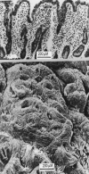
The SEM reveals that the surface openings (see Fig 1) are large crevices up to 200mM in length. Within the depths of some of these crevices, the openings of individual crypt tubes are visible. Each crevice (basin) is surrounded by concentric arrays of enterocytes (“collars”). Some collars are elevated above the plane of flattening.
Figure 3
Figure 3
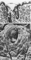
Close-up of another individual basin and surrounding crypt collars. Above, a corresponding histological view of the same specimen, showing two individual crypt tubes opening into the basin. On left, the section through the collar could be mistaken for a “villous” projection.
Figure 4
Figure 4
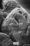
Closer view of a single crypt basin. White arrow heads locate openings of individual crypt tubes at bottom of basin. M, mucus blob.
Figure 5
Figure 5
SEM appearances of a small mosaic plateau bearing the large openings of several basins. Some may be seen to be perforated in their depths by individual crypt tubes. Other openings of individual crypt tubes are visible at the base of the adjacent furrow to the left of the plateau illustrated.
Figure 6
Figure 6
This specimen, obtained after 5 months of dietary gluten restriction, reveals two elevated, adjacent collars that appear to have joined to form a curvilinear ridge. These ridges will ultimately thicken to form low ‘convolutions’ from which true villous projections will ultimately take origin, although at a much later stage in mucosal regeneration. On the right, imaginary appearances of random sections across this specimen are shown. One (A) reveals the result of a section along the flat surface contour relevant to the conjoined ridge. The other (B), passing through the collared basin, reveals pseudo-villous contours, even though overall, the specimen is still flat. This is a prime example of how the surface contour varies within a micro-region across every specimen, and clearly demonstrating that any random, thin histological section cannot provide useful information about surface shape and contour.

References

    1. Marsh MN. Gluten, major histocompatibility complex, and the small intestine: A molecular and immunobiologic approach to the spectrum of gluten sensitivity (‘celiac sprue’) Gastroenterology. 1992;102:330–54. - PubMed
    1. Egan-Mitchell B, Fottrell PF, McNichol BF. Early or pre-celiac mucosa: development of gluten enteropathy. Gut. 1981;22:65–69. - PMC - PubMed
    1. Marsh MN. Studies of intestinal lymphoid tissue: XIII – Immunopathology of the evolving celiac sprue lesion. Pathol Res Pract. 1989;185:774–77. - PubMed
    1. Maki M, Holm K, Koskimies S, Hallstrom O, Visakorpi J. Normal small bowel biopsy followed by coeliac disease. Arch Dis Child. 1990;65:1137–41. - PMC - PubMed
    1. Paulley JW. Observations on the aetiology of idiopathic steatorrhoea. Br Med J. 1954:1318–21. - PMC - PubMed

LinkOut - more resources