. 2015 Apr 29:3:5.
doi: 10.1186/s40170-015-0131-7. eCollection 2015.

An epitope tag alters phosphoglycerate dehydrogenase structure and impairs ability to support cell proliferation

Affiliations

An epitope tag alters phosphoglycerate dehydrogenase structure and impairs ability to support cell proliferation

Katherine R Mattaini et al. Cancer Metab. .

Abstract

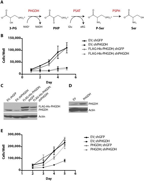
Background: The gene encoding the serine biosynthesis pathway enzyme PHGDH is located in a region of focal genomic copy number gain in human cancers. Cells with PHGDH amplification are dependent on enzyme expression for proliferation. However, dependence on increased PHGDH expression extends beyond production of serine alone, and further studies of PHGDH function are necessary to elucidate its role in cancer cells. These studies will require a physiologically relevant form of the enzyme for experiments using engineered cell lines and recombinant protein.

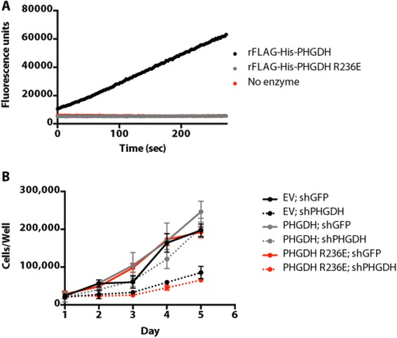
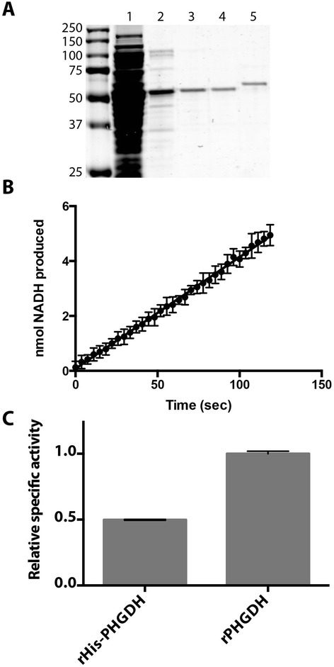
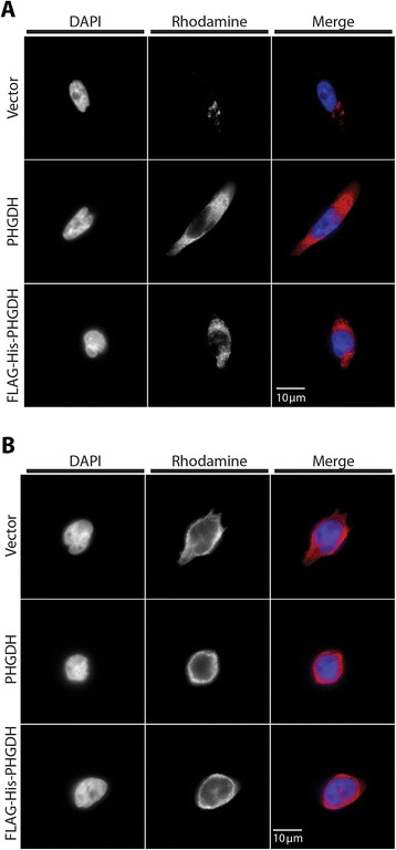
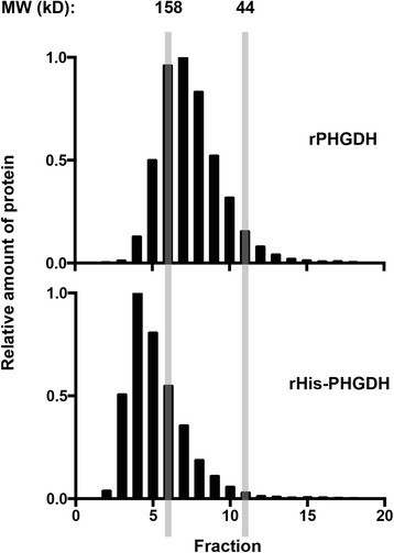
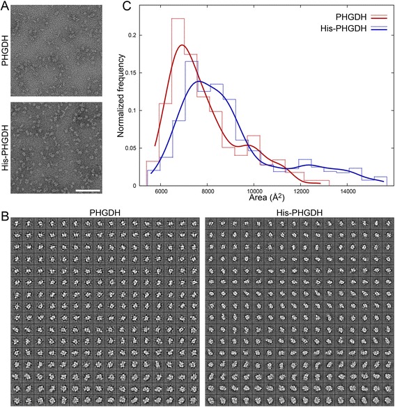
Results: The addition of an N-terminal epitope tag to PHGDH abolished the ability to support proliferation of PHGDH-amplified cells despite retention of some activity to convert 3-PG to PHP. Introducing an R236E mutation into PHGDH eliminates enzyme activity, and this catalytically inactive enzyme cannot support proliferation of PHGDH-dependent cells, arguing that canonical enzyme activity is required. Tagged and untagged PHGDH exhibit the same intracellular localization and ability to produce D-2-hydroxyglutarate (D-2HG), an error product of PHGDH, arguing that neither mislocalization nor loss of D-2HG production explains the inability of epitope-tagged PHGDH to support proliferation. To enable studies of PHGDH function, we report a method to purify recombinant PHGDH and found that untagged enzyme activity was greater than N-terminally tagged enzyme. Analysis of tagged and untagged PHGDH using size exclusion chromatography and electron microscopy found that an N-terminal epitope tag alters enzyme structure.

Conclusions: Purification of untagged recombinant PHGDH eliminates the need to use an epitope tag for enzyme studies. Furthermore, while tagged PHGDH retains some ability to convert 3PG to PHP, the structural alterations caused by including an epitope tag disrupts the ability of PHGDH to sustain cancer cell proliferation.

Keywords: Cancer Metabolism; Epitope tag; Phosphoglycerate dehydrogenase; Serine metabolism; Serine synthesis.


Figures

Figure 1
Figure 1
N-terminal epitope-tagged PHGDH cannot support cell proliferation following PHGDH knockdown. (A) Schematic representation of the serine biosynthesis pathway. 3-PG, 3-phosphoglycerate; PHGDH, 3-phosphoglycerate dehydrogenase; NAD+/NADH, oxidized and reduced forms of nicotinamide adenine dinucleotide, respectively; PHP, phosphohydroxypyruvate; PSAT, phosphoserine aminotransferase; Glu, glutamate; αKG, alpha-ketoglutarate; P-Ser, phosphoserine; PSPH, phosphoserine phosphatase; Ser, serine. (B) Cell number over time of PHGDH-amplified T.T. cells stably expressing either an shRNA-resistant FLAG-His-PHGDH cDNA or empty vector (EV) control. (C) Western blot analysis assessing knockdown of endogenous PHGDH and expression of FLAG-His-PHGDH cDNA. (D) Western blot analysis of T.T. cells stably expressing an shRNA-resistant PHGDH cDNA (untagged) or empty vector (EV) control. (E) Cell number over time of the cells described in (D) when infected with virus expressing GFP or PHGDH shRNA. Error bars show standard deviation from the mean.
Figure 2
Figure 2
PHGDH enzymatic activity is required for cell proliferation following PHGDH knockdown. (A) In vitro enzyme activity assessed by tracking NADH production by fluorescence. The assay was performed at saturating substrate concentrations. (B) Cell number over time for PHGDH-amplified T.T. cells stably expressing shRNA-resistant PHGDH wild type or R236E (enzymatically dead) cDNAs or empty vector (EV) control when infected with virus expressing GFP or PHGDH shRNA. Error bars show standard deviation from the mean.
Figure 3
Figure 3
Purified untagged recombinant PHGDH has higher specific activity than N-terminally tagged PHGDH. (A) Coomassie gel of rPHGDH (untagged) after each step of the purification: 1. cleared lysate from E. coli expressing rPHGDH protein, 2. eluate from Cibacron Blue F3GA column, 3. eluate from anion exchange column, 4. eluate from size exclusion column, and 5. rFLAG-His-PHGDH purified by sequential nickel and size exclusion columns, for comparison. Molecular weight standards in kilodaltons are present to the left of lane 1. (B) In vitro enzyme activity of untagged rPHGDH. Assay was performed at saturating substrate concentrations. (C) Comparison of relative specific activity of rPHGDH and rHis-PHGDH. Error bars show standard deviation from the mean.
Figure 4
Figure 4
N-terminal epitope-tagged and untagged PHGDH display the same intracellular localization. (A) MDA-MB-231 cells, which are PHGDH low, and (B) PHGDH-amplified T.T. cells expressing either vector control, untagged, or N-terminally tagged PHGDH were stained for PHGDH immunofluorescence.
Figure 5
Figure 5
N-terminal epitope-tagged and untagged PHGDH exhibit different mobility on size exclusion chromatography. Relative amount of rPHGDH and rHis-PHGDH protein present in fractions eluted from a Sephacryl S-200 size exclusion column. The 158- and 44-kDa standards are shown for comparison, with the column void volume represented in fraction 2.
Figure 6
Figure 6
N-terminally tagged PHGDH forms larger macromolecular complexes than untagged PHGDH. (A) Representative images obtained from PHGDH and His-PHGDH specimens. Scale bar = 100 nm. (B) Montage of class averages of PHGDH and His-PHGDH sorted from smallest (top row) to largest (bottom row). Each average is 294 Å wide at the specimen level. (C) Normalized particle-size distributions (particles per bin/total particles) are displayed as histograms (dashed lines) and as smoothed curves (solid lines) for PHGDH (blue) and His-PHGDH (red). The His-PHGDH and PHGDH histograms are offset by 42 Å2 to avoid overlapping lines.

References

    1. Locasale JW. Serine, glycine and one-carbon units: cancer metabolism in full circle. Nat Rev Cancer. 2013;13:572. doi: 10.1038/nrc3557. - DOI - PMC - PubMed
    1. Mullarky E, Mattaini KR, Vander Heiden MG, Cantley LC, Locasale JW. PHGDH amplification and altered glucose metabolism in human melanoma. Pigment Cell Melanoma Res. 2011;24:1112. doi: 10.1111/j.1755-148X.2011.00919.x. - DOI - PubMed
    1. Locasale JW, Grassian AR, Melman T, Lyssiotis CA, Mattaini KR, Bass AJ, et al. Phosphoglycerate dehydrogenase diverts glycolytic flux and contributes to oncogenesis. Nat Genet. 2011;43:869. doi: 10.1038/ng.890. - DOI - PMC - PubMed
    1. Possemato R, Marks KM, Shaul YD, Pacold ME, Kim D, Birsoy K, et al. Functional genomics reveal that the serine synthesis pathway is essential in breast cancer. Nature. 2011;476:346. doi: 10.1038/nature10350. - DOI - PMC - PubMed
    1. Nilsson LM, Forshell TZP, Rimpi S, Kreutzer C, Pretsch W, Bornkamm GW, et al. Mouse genetics suggests cell-context dependency for Myc-regulated metabolic enzymes during tumorigenesis. PLoS Genet. 2012;8:e1002573. doi: 10.1371/journal.pgen.1002573. - DOI - PMC - PubMed