

Raman spectroscopic sensing of carbonate intercalation in breast microcalcifications at stereotactic biopsy
- PMID: 25927331
- PMCID: PMC4415591
- DOI: 10.1038/srep09907
Raman spectroscopic sensing of carbonate intercalation in breast microcalcifications at stereotactic biopsy
Abstract
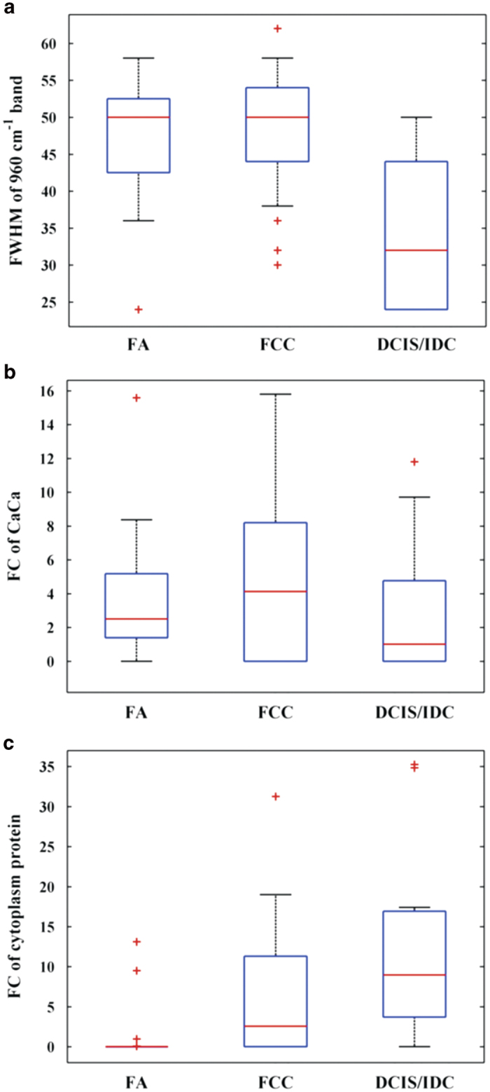
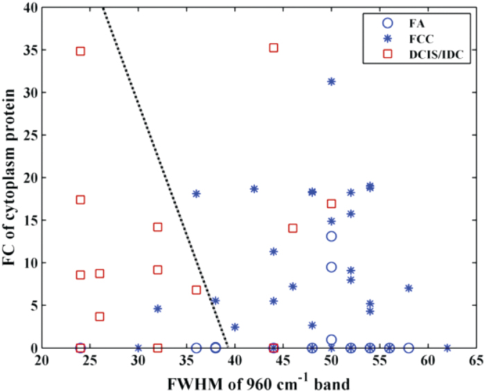
Microcalcifications are an early mammographic sign of breast cancer and frequent target for stereotactic biopsy. Despite their indisputable value, microcalcifications, particularly of the type II variety that are comprised of calcium hydroxyapatite deposits, remain one of the least understood disease markers. Here we employed Raman spectroscopy to elucidate the relationship between pathogenicity of breast lesions in fresh biopsy cores and composition of type II microcalcifications. Using a chemometric model of chemical-morphological constituents, acquired Raman spectra were translated to characterize chemical makeup of the lesions. We find that increase in carbonate intercalation in the hydroxyapatite lattice can be reliably employed to differentiate benign from malignant lesions, with algorithms based only on carbonate and cytoplasmic protein content exhibiting excellent negative predictive value (93-98%). Our findings highlight the importance of calcium carbonate, an underrated constituent of microcalcifications, as a spectroscopic marker in breast pathology evaluation and pave the way for improved biopsy guidance.
Figures

References
-
- Department of Defense Breast Cancer Research Program, The Breast Cancer Landscape. Technical Report. (2013) Available at: http://cdmrp.army.mil/bcrp/pdfs/bc_landscape13.pdf (Accessed: 12th February 2015).
-
- Tabar L. et al. A novel method for prediction of long-term outcome of women with T1a, T1b, and 10–14 mm invasive breast cancers: A prospective study. Lancet. 355, 429–33 (2000). - PubMed
-
- Evans A. J. et al. Mammographic features of ductal carcinoma in situ (DCIS) present on previous mammography. Clin. Radiol. 54, 644–646 (1999). - PubMed
-
- Johnson J. et al. Histological correlation of microcalcifications in breast biopsy specimens. Arch. Surg. 134, 712–715 (1999). - PubMed
-
- Gulsun M., Demirkazik F. B. & Ariyurek M. Evaluation of breast microcalcifications according to Breast Imaging Reporting and Data System criteria and Le Gal’s classification. Eur. J. Radiol. 47, 227–231 (2003). - PubMed
Publication types
MeSH terms
Substances
Grants and funding
LinkOut - more resources
Full Text Sources
Other Literature Sources
