. 2015;14(11):1716-29.
doi: 10.1080/15384101.2015.1033595.

PRMT5- mediated symmetric arginine dimethylation is attenuated by mutant huntingtin and is impaired in Huntington's disease (HD)

Affiliations

PRMT5- mediated symmetric arginine dimethylation is attenuated by mutant huntingtin and is impaired in Huntington's disease (HD)

Tamara Ratovitski et al. Cell Cycle. 2015.

Abstract

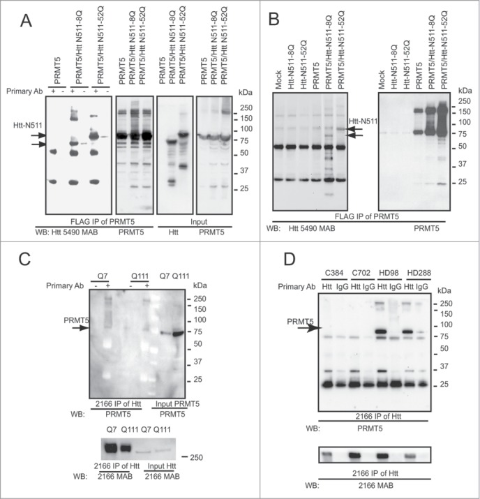
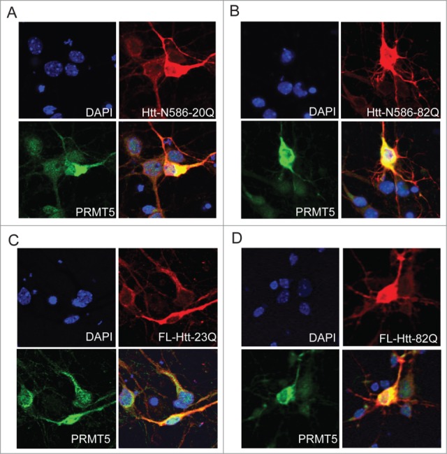
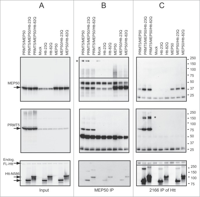
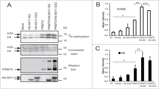
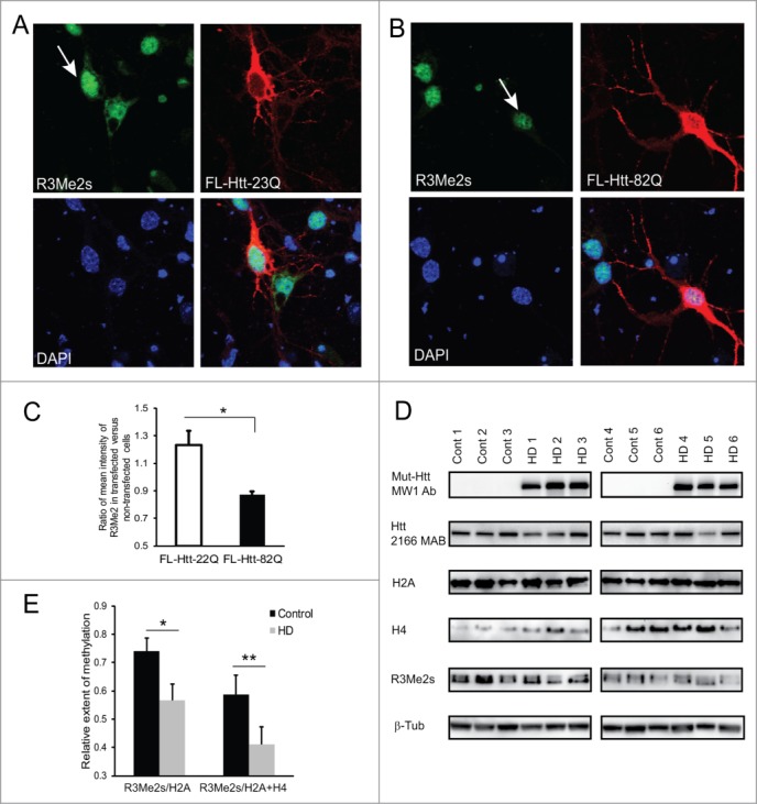
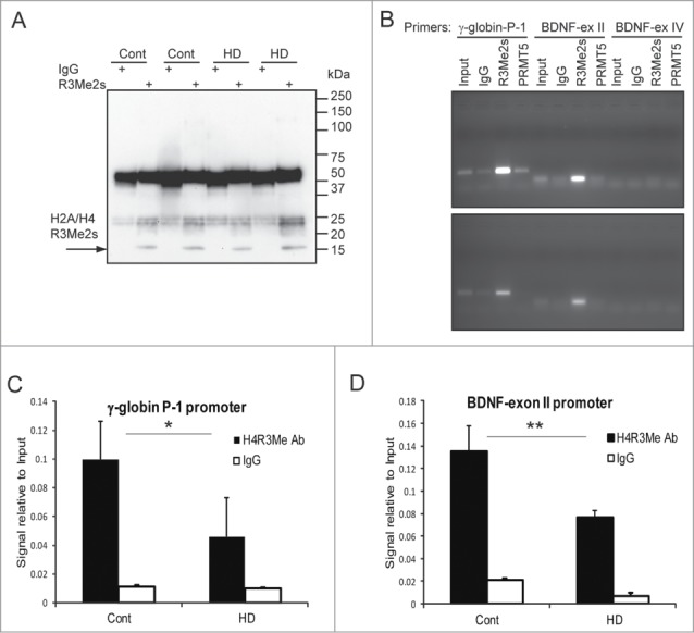
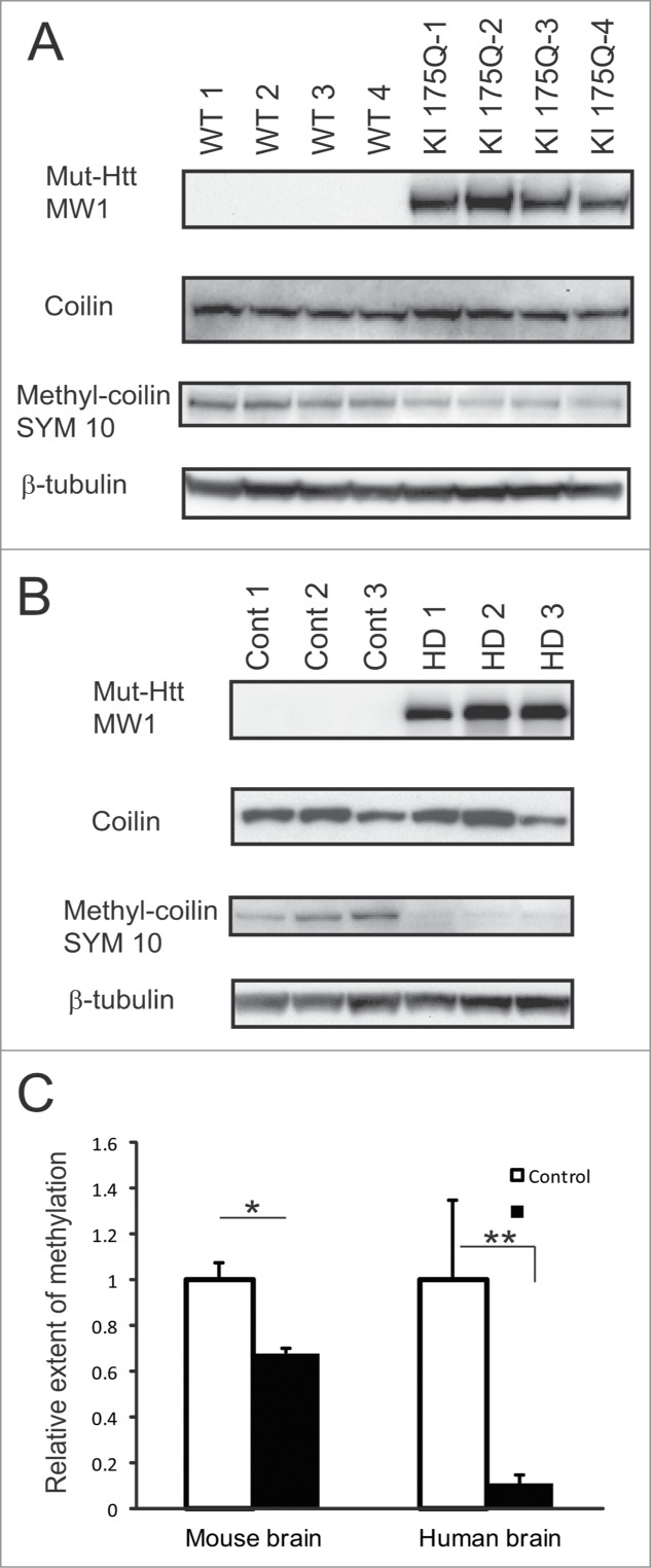
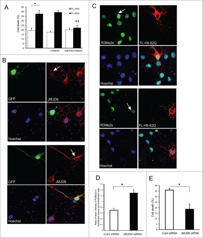
Abnormal protein interactions of mutant huntingtin (Htt) triggered by polyglutamine expansion are thought to mediate Huntington's disease (HD) pathogenesis. Here, we explored a functional interaction of Htt with protein arginine methyltransferase 5 (PRMT5), an enzyme mediating symmetrical dimethylation of arginine (sDMA) of key cellular proteins, including histones, and spliceosomal Sm proteins. Gene transcription and RNA splicing are impaired in HD. We demonstrated PRMT5 and Htt interaction and their co-localization in transfected neurons and in HD brain. As a result of this interaction, normal (but to a lesser extend mutant) Htt stimulated PRMT5 activity in vitro. SDMA of histones H2A and H4 was reduced in the presence of mutant Htt in primary cultured neurons and in HD brain, consistent with a demonstrated reduction in R3Me2s occupancy at the transcriptionally repressed promoters in HD brain. SDMA of another PRMT5 substrate, Cajal body marker coilin, was also reduced in the HD mouse model and in human HD brain. Finally, compensation of PRMT5 deficiency by ectopic expression of PRMT5/MEP50 complexes, or by the knock-down of H4R3Me2 demethylase JMJD6, reversed the toxic effects of mutant Htt in primary cortical neurons, suggesting that PRMT5 deficiency may mediate, at least in part, HD pathogenesis. These studies revealed a potential new mechanism for disruption of gene expression and RNA processing in HD, involving a loss of normal function of Htt in facilitation of PRMT5, supporting the idea that epigenetic regulation of gene transcription may be involved in HD and highlighting symmetric dimethylation of arginine as potential new therapeutic target.

Keywords: BDNF, brain-derived neurotrophic factor; CB, Cajal body; ChIP, the chromatin immunoprecipitation; DMEM, Dulbecco's modified Eagle's medium; FBS, fetal bovine serum; HD, Huntington's disease; HEK, human embryonic kidney; Htt, huntingtin; Huntington's disease mechanism; IP, immunoprecipitation; IgG, immunoglobulin; PIC, protease inhibitors cocktail; PRMT5, protein arginine methyltransferase; RNA processing; SMN, survival of motor neurons; Sm proteins, spleceosomal small nuclear ribonucleoproteins; gene transcription; huntingtin; neurodegeneration; polyQ, polyglutamine; protein interactions; protein methylation; sDMA, symmetrical arginine dimethylation; snRNPs, small nuclear ribonucleoprotein particles.


Figures

Figure 1.
Figure 1.
Htt interacts with PRMT5 in cell lines and human brain. (A) HEK 293 cells were transiently transfected with the indicated plasmids, keeping the amount of Htt and PRMT5 plasmids and total amount of DNA equal between transfections by supplementing with empty vector. Cells were lysed 48 h after transfection, and PRMT5 complexes were immunoprecipitated using the anti-FLAG-agarose beads. Normal and expanded Htt were detected in the immunoprecipitates (IPs) from cells co-transfected with Htt and PRMT5, but not in control samples without the primary antibody or in cells transfected with Htt constructs only (first panel and data not shown). IPs were also analyzed for the presence of PRMT5 using the PRMT5-specific antibody (second panel). The inputs are shown on the right 2 panels. (B) Rat striatal ST14 cells were transiently transfected with indicated plasmids, lysed 48 h after transfection, and PRMT5 complexes were immunoprecipitated using the anti-FLAG-agarose beads. Normal and expanded Htt were detected in the IPs from cells co-transfected with Htt and PRMT5, but not in mock transfection, or in cells transfected with Htt constructs only (left panel). IPs were also analyzed for the presence of PRMT5 using PRMT5-specific antibody (right panel). (C) Endogenous Htt proteins were immunoprecipitated from STHdh Q7/Q7 and Q111/Q111 cells using a specific antibody to Htt (MAB2166). Endogenous PRMT5 monomers and high-molecular weight PRMT5 complexes were detected in the IPs using the PRMT5-specific antibody, but not in control samples without the primary antibody (top panel, left side). PRMT5 inputs are shown on the last 2 lanes. IPs were also analyzed for the presence of Htt using the Htt-specific antibody, and Htt inputs are shown (bottom panel). (D) Htt complexes were immunoprecipitated from total cell homogenates from frozen human front superior cortex of normal controls and HD cases. Endogenous PRMT5 monomers and high-molecular weight PRMT5 complexes were detected in the IPs from HD brain using the PRMT5-specific antibody, but not in normal controls or in control samples where normal IgGs were used for IP (top panel). Htt inputs are shown on the bottom panel.
Figure 2.
Figure 2.
Htt and PRMT5 co-localize in primary neurons. Primary mouse cortical neurons were co-transfected at 5 DIV with the expression vectors for PRMT5 and either normal or polyQ-expanded Htt N586 fragment (A, B) or the full-length Htt constructs (C, D). Confocal immunofluorescence detection of Htt with 2166 monoclonal antibody is shown in red (Alexa Fluor 555); detection of PRMT5 with polyclonal specific antibody is shown in green (Alexa Fluor 488); nuclear staining (DAPI) is shown in blue. Yellow staining in merged images demonstrated partial co-localization. Representative images are shown for each construct.
Figure 3.
Figure 3.
Htt interacts with PRMT5/MEP50 complex. HEK 293 cells were transiently transfected with the indicated plasmids, keeping the amount of Htt-N586, PRMT5 and MEP50 plasmids and total amount of DNA equal between transfections by supplementing with empty vector. Cells were lysed 48 h after transfection, and MEP50 (B) or Htt (C) complexes were immunoprecipitated using either MEP50-or Htt-specific antibodies. All three proteins-MEP50 (top panels), PRMT5 (middle panels) and Htt (bottom panels) were detected in both MEP50 and Htt IPs from cells co-transfected with Htt, PRMT5 and MEP50. High-molecular weight complexes detected with PRMT5 and MEP50 antibodies are marked by asterisk. The inputs are shown in A.
Figure 4.
Figure 4.
Htt modulates PRMT5 activity toward histones. (A) In vitro methyltransferase assay of FLAG IPs from ST14 cells transfected with indicated plasmids, keeping the amount of Htt and PRMT5 plasmids and total amount of DNA equal between transfections by supplementing with empty vector. Autoradiographs (upper panel), Coomassie stained gel (middle panel) are shown. Western blot detections of PRMT5 with PRMT5-specific antibody and of Htt with MAB5490 antibody are shown on bottom panels. (B, C) The graphs show the quantification of autoradiographs from 3 experiments for histones H2A (B) and H4 (C), *n = 3, p = 0.005; **n = 3, p = 0.02; ***n = 3, p = 0.1.
Figure 5.
Figure 5.
Symmetric arginine dimethylation of histones is attenuated by mutant Htt in primary neurons and is impaired in HD brain. (A, B) Primary mouse cortical neurons were transfected at 5 DIV with normal (A) or mutant (B) full-length Htt expression constructs. Representative images of confocal immunofluorescence detection of Htt with 2166 monoclonal antibody (in red, Alexa Fluor 555) and of sDMA modification of H2A and H4 with R3Me2s modification-specific antibody (in green, Alexa Fluor 488) are shown. Transfected cells are indicated with white arrows The nuclear staining (DAPI) is shown in blue. (C) Quantification of H4R3me2s staining in transfected cells presented as a ratio of mean intensity of the staining in transfected and non-transfected cells. 100 transfected cells were analyzed for each condition in 3 experiments. (*n = 3, p = 0.004). (D) Total cell homogenates from frozen human front superior cortex of 6 normal controls and 6 HD cases were analyzed by Western blotting using the following antibodies: MW1 for detection of expanded Htt; 2166 MAB for detection of normal and expanded Htt; histone-specific antibodies for detection of histones H2A and H4; modification-specific antibody R3Me2s for detection of sDMA on R3 of histones H2A and H4; β-tubulin as a loading control. Representative blots are shown. (E) Quantification of the levels of R3Me2s modification on histones H2A and H4 presented as a ratio of R3Me2s signal intensity to total levels of histones based on Western blot repeated 3 times (*n = 3, p = 0.045; ** n = 3, p = 0.045).
Figure 6.
Figure 6.
Reduction of H2A/H4R3Me2s occupancy at the γ-globin and BDNF promoters in HD brain. (A) Confirmation of ChIP assay by Western blotting with the R3Me2s modification-specific antibody. Representative image for 2 controls and 2 HD samples is shown. (B) Detection of H2A/H4 R3Me2s marks at the γ-globin P-1 and BDNF promoters by ChIP assay. Chromatin fractions from HD (bottom panel) or control (top panel) human brain were immunoprecipitated with the indicated antibodies and amplified with the indicated primer pairs. Normal rabbit IgG used as a negative control. (C and D) R3Me2s enrichment at the γ-globin P-1 (C) and BDNF-ex II promoters (D) were measured by ChIP and q-PCR assays in HD and control human brains (*n = 4, p = 0.1; **n = 3, p = 0.1).
Figure 7.
Figure 7.
Coilin symmetric dimethylation is reduced in HD mouse and human brain. (A, B) Whole brain homogenates from 4 KI175Q HD and 4 WT mice (A) or homogenates from frozen human frontal superior cortex of 5 normal controls and 5 HD cases (B) were analyzed by Western blotting using the following antibodies: MW1 for detection of expanded Htt; coilin-specific antibody; SYM10 for detection of symmetrically dimethylated coilin and β-tubulin as a loading control. Representative images are shown. (C) Quantification of the relative extent of symmetric dimethylation of coilin presented as a ratio of SYM10 signal intensity to total levels of coilin. (*n = 4, p = 0.007; **n = 5, p = 0.03).The experiment was repeated 3 times.
Figure 8.
Figure 8.
Modulation of the levels of arginine methyltransferase PRMT5 and histone arginine demethylase JMJD6 affects the survival of HD neurons. (A) Co-transfection of PRMT5 with its cofactor MEP50 rescued primary neurons from the toxicity of mutant Htt. Primary cortical neurons were co-transfected at 5 DIV with either normal or mutant full-length Htt and PRMT5 and MEP50 expression plasmids as indicated, and co-transfected with eGFP to identify transfected cells. Cell death was measured by nuclear condensation assay. * n = 4, P < 0.01; ** (co-transfected with PRMT5 and MEP50 vs with Htt alone) n = 4, P < 0.01. (B–D) JMJD6 knock-down rescued primary neurons from the toxicity of mutant Htt. (B) Primary mouse cortical neurons were co-transfected at 5 DIV with mutant full-length Htt expression constructs, eGFP (to identify transfected cells) and either JMJD6 siRNA (top panels) or with non-targeting control siRNA (bottom panels). Representative images of confocal immunofluorescence detection of JMJD6 (in red, Alexa Fluor 555) in GFP-positive (transfected, indicated with white arrow) and GFP-negative (non-transfected) cells are shown. The nuclear staining (Hoechst) is shown in blue. (C) Primary mouse cortical neurons were co-transfected at 5 DIV with mutant full-length Htt expression constructs and either JMJD6 siRNA (top panels) or with non-targeting control siRNA (bottom panels). Representative images of confocal immunofluorescence detection of Htt with 2166 monoclonal antibody (in red, Alexa Fluor 555) and of sDMA modification of H2A and H4 with R3Me2s modification-specific antibody (in green, Alexa Fluor 488) in transfected (indicated with white arrow) and non-transfected cells are shown. The nuclear staining (Hoechst) is shown in blue. (D) Quantification of H4R3Me2s staining in transfected cells presented as a ratio of mean intensity of the staining in transfected and non-transfected cells. 100 transfected cells were analyzed for each condition in 3 experiments. (*n = 3, P < 0.001). (E) Primary cortical neurons were co-transfected with mutant full-length Htt-82Q and either JMJD6 siRNA or with non-targeting control siRNA as indicated, and co-transfected with eGFP to identify transfected cells. Cell death was measured by nuclear condensation assay. ˜300 cells were analyzed per each condition in each experiment. * n = 3, P < 0.001

Similar articles

Cited by

References

    1. A novel gene containing a trinucleotide repeat that is expanded and unstable on Huntington's disease chromosomes . The Huntington's Disease Collaborative Research Group. Cell 1993; 72:971-83; PMID:8458085; http://dx.doi.org/10.1016/0092-8674(93)90585-E - DOI - PubMed
    1. Ross CA, Aylward EH, Wild EJ, Langbehn DR, Long JD, Warner JH, Scahill RI, Leavitt BR, Stout JC, Paulsen JS, et al.. Huntington disease: natural history, biomarkers and prospects for therapeutics. Nat Rev Neurol 2014; 10:204-16; PMID:24614516; http://dx.doi.org/10.1038/nrneurol.2014.24 - DOI - PubMed
    1. Ross CA, Tabrizi SJ. Huntington's disease: from molecular pathogenesis to clinical treatment. Lancet Neurol 2011; 10:83-98; PMID:21163446; http://dx.doi.org/10.1016/S1474-4422(10)70245-3 - DOI - PubMed
    1. Walker FO . Huntington's disease. Lancet 2007; 369:218-28; PMID:17240289; http://dx.doi.org/10.1016/S0140-6736(07)60111-1 - DOI - PubMed
    1. Boutell JM, Thomas P, Neal JW, Weston VJ, Duce J, Harper PS, Jones AL. Aberrant interactions of transcriptional repressor proteins with the Huntington's disease gene product, huntingtin. Hum Mol Genet 1999; 8:1647-55; PMID:10441327; http://dx.doi.org/10.1093/hmg/8.9.1647 - DOI - PubMed

Publication types