. 2015 Apr 30;10(4):e0124657.
doi: 10.1371/journal.pone.0124657. eCollection 2015.

The shh signaling pathway is upregulated in multiple cell types in cortical ischemia and influences the outcome of stroke in an animal model

Affiliations

The shh signaling pathway is upregulated in multiple cell types in cortical ischemia and influences the outcome of stroke in an animal model

Yongmin Jin et al. PLoS One. .

Abstract

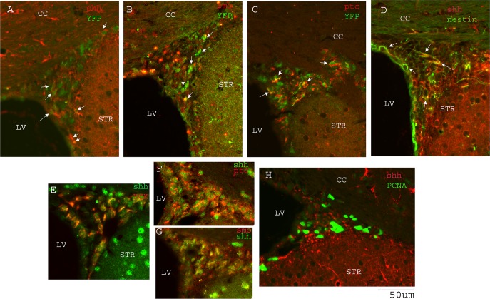
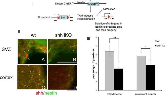
Recently the sonic hedgehog (shh) signaling pathway has been shown to play an important role in regulating repair and regenerative responses after brain injury, including ischemia. However, the precise cellular components that express and upregulate the shh gene and the cellular components that respond to shh signaling remain to be identified. In this study, using a distal MCA occlusion model, our data show that the shh signal is upregulated both at the cortical area near the injury site and in the adjacent striatum. Multiple cell types upregulate shh signaling in ischemic brain, including neurons, reactive astrocytes and nestin-expressing cells. The shh signaling pathway genes are also expressed in the neural stem cells (NSCs) niche in the subventricular zone (SVZ). Conditional deletion of the shh gene in nestin-expressing cells both at the SVZ niche and at the ischemic site lead to significantly more severe behavioral deficits in these shh iKO mice after cortical stroke, measured using an automated open field locomotion apparatus (Student's t-test, p<0.05). In contrast, animals given post-stroke treatment with the shh signaling agonist (SAG) demonstrated less deficits in behavioral function, compared to vehicle-treated mice. At 7 days after stroke, SAG-treated mice showed higher values in multiple horizontal movement parameters compared to vehicle treated mice (Student's t-test, p<0.05) whereas there were no differences in pre-stroke measurements, (Student's t-test, p>0.05). In summary, our data demonstrate that shh signaling plays critical and ongoing roles in response to ischemic injury and modulation of shh signaling in vivo alters the functional outcome after cortical ischemic injury.


Conflict of interest statement

Competing Interests: The authors have declared that no competing interests exist.

Figures

Fig 1
Fig 1. Expression of yellow fluorescent protein (YFP) and double cortin (DCX) in neural progenitor/neuroblast cells in stroke brain.
A) experimental timeline. B) TTC staining at 48 hours after distal MCAo showing infarction restricted to cortical area. C) MAP2 immunostaining reveals loss of neurons in cortical stroke D) and E) YFP and DCX immunostaining in SVZ in the contralateral and ipsilateral sides of the brain. Inset shows the staining at higher magnification.. F) quantification of SVZ YFP+/DCX+ cells in the contralateral and ipsilateral side of the brain in distal MCAo mice (n = 6). G) TTC staining showing a representative image of infarction in proximal MCAo H) post-stroke day (psd 30) DCX immunostaining showing minimal migration of DCX+ positive cells into the ischemic site in distal MCAo. Boxed area in (H) is shown in panel I) at higher magnification. J) shows many migrating DCX+ neuroblasts toward the ischemic injury site in proximal MCAo. Boxed area in (J) is shown in panel K) at higher magnification. L) Quantification of total DCX positive cells per section at the ischemic sites in distal MCAo and proximal MCAo mice (n = 3–4 per group). Scale bar = 100um. ** indicates p<0.01, Student’s t-test.
Fig 2
Fig 2. Distal MCAo induces shh expression in the cortical ischemic site.
A) and C) expression of shh and MAP2 in the contralateral side cortex. B) and D) shh expression in the ischemic site of the ipsilateral cortex. Arrow and inset showing a few shh+/MAP2+ cells. E) basal expression of shh and minimal or no expression of nestin in contralateral side of cortex and F) shh and nestin upregulation near the ischemic site in ipsilateral cortex. G and H) upregulation of GFAP in both cortex and striatum in the ipsilateral side. I and J) expression of shh and GFAP in the contralateral and ipsilateral sides of cortex at higher magnification. K/L/M) staining of nestin/GFAP/shh on the same section. N) Quantification of immunofluorescent intensity for shh, nestin and GFAP in contralateral and ipsilateral sides of the cortical area (n = 6). O, P, Q and R) colocalization analysis of shh/MAP2, shh/GFAP, shh/nestin and nesin/GFAP signals (protein on the y axis to the protein on x axis as labeled on the scatterplots. Pearson’s correlation coefficient (PCC) are marked on the bottom of the scatterplots. Scale bar = 100um. ** indicates p<0.01, Student’s t-test. Mouse brains were analyzed at 14 days after distal MCAo.
Fig 3
Fig 3. Upregulation of shh expression in the ipsilateral striatum in GFAP positive cells.
A) Basal expression of shh in the contralateral striatum in GFAP negative cells, C) showing higher magnification. Shh and GFAP are upregulated in striatum in cortical distal MCAo model B). Higher magnification of shh+/GFAP+ cells are shown in panel D). E) Quantification of immunofluorescent intensity for shh and GFAP in contralateral and ipsilateral sides of the adjacent striatal area (n = 6). F) percentage of shh+ cells that are GFAP+ or nestin + in ipsilateral striatal area (>100 total cells counted). Scale bar is as marked. * indicates p<0.05 and ** indicates p<0.01, Student’s t-test. Mouse brains were analyzed at 14 days after distal MCAo.
Fig 4
Fig 4. Nestin expression is induced in ipsilateral striatum.
A.B.C) basal expression of shh and minimal or no expression of nestin protein in the contralateral striatum. D) nestin expression is upregulated in the ipsilateral striatum and many of the nestin+ cells are also shh+ (E and F). Higher magnification image is shown in panel G. (H) Some of the nestin-expressing cells in striatum are also GFAP positive. Scale bar is as marked. Mouse brains were analyzed at 14 days after distal MCAo.
Fig 5
Fig 5. Shh signaling pathway genes are expressed in SVZ NSCs.
A.B.C) shh, smo, ptc expression in YFP (+) cells in the SVZ. D) shh expression in nestin(+) NSCs in SVZ. E). shh expression is detected in DCX+ neuroblasts in SVZ. H) shh expression in PCNA(+) cells. F.G) coexpression of shh with smo, and shh with ptc in SVZ cells. Scale bar = 50um. Mouse brains were analyzed at 14 days after distal MCAo.
Fig 6
Fig 6. Nestin(+) cell-specific deletion of shh gene leads to more motor function deficits in shh iKO mice.
I) strategy for nestin-expressing cell specific deletion of shh. II) in wt mice, shh expression is detected in many cells that are also nestin(+) in the SVZ (A) and in the ischemic site of the cortical stroke area (C). However, in shh iKO mice, only very few nestin positive cells express shh (panel B arrow). Similarly at the ischemic site in cortex (D), nestin (+) cells do not express shh while surrounding cells expressing shh are nestin negative. This confirms the deletion of the shh gene in both SVZ and cortical nestin-expressing cells near the ischemic site. III) Deletion of shh gene in nestin(+) cells in shh iKO mice leads to greater motor deficits in these ko mice compared to wt mice measured by total distance travelled and total movement number recorded in 24 hours at post-stroke day 7. Data normalized to pre-stroke value of each animal and presented as percentage of pre-stroke measurements. (** indicates p<0.01, * indicates p<0.05, n = 7–8, Student’s t-test). Scale bar = 50um.
Fig 7
Fig 7. Shh pathway agonist treatment starting at post-stroke day (psd 3) improves motor function in stroke mice.
Motor function was measured by total distance travelled (cm) and total movement number recorded in 24 hours. Measurement was carried out before MCAo surgery and on post-stroke day 7. (n = 15, * indicates p<0.05, Student’s t-test).

References

    1. Gilman S. Pharmacologic management of ischemic stroke: relevance to stem cell therapy. Exp Neurol. 2006;199(1):28–36. . - PubMed
    1. Goldstein LB. Acute ischemic stroke treatment in 2007. Circulation. 2007;116(13):1504–14. . - PubMed
    1. Luo Y. Cell-based therapy for stroke. Journal of neural transmission. 2011;118(1):61–74. 10.1007/s00702-010-0478-4 . - DOI - PubMed
    1. Magnusson JP, Goritz C, Tatarishvili J, Dias DO, Smith EM, Lindvall O, et al. A latent neurogenic program in astrocytes regulated by Notch signaling in the mouse. Science. 2014;346(6206):237–41. 10.1126/science.346.6206.237 . - DOI - PubMed
    1. Ruiz i Altaba A, Palma V, Dahmane N. Hedgehog-Gli signalling and the growth of the brain. Nat Rev Neurosci. 2002;3(1):24–33. Epub 2002/02/02. 10.1038/nrn704nrn704 [pii]. . - DOI - PubMed

Publication types