

Modulation of bleomycin-induced lung fibrosis by pegylated hyaluronidase and dopamine receptor antagonist in mice
- PMID: 25927611
- PMCID: PMC4415936
- DOI: 10.1371/journal.pone.0125065
Modulation of bleomycin-induced lung fibrosis by pegylated hyaluronidase and dopamine receptor antagonist in mice
Abstract
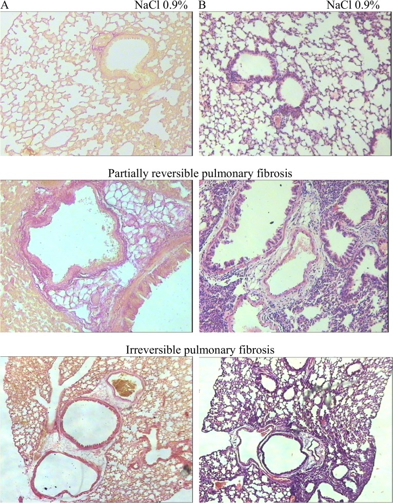
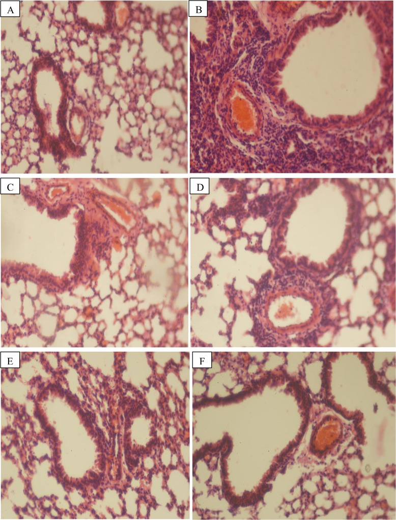
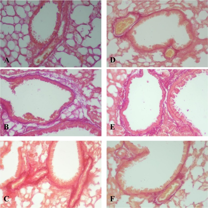
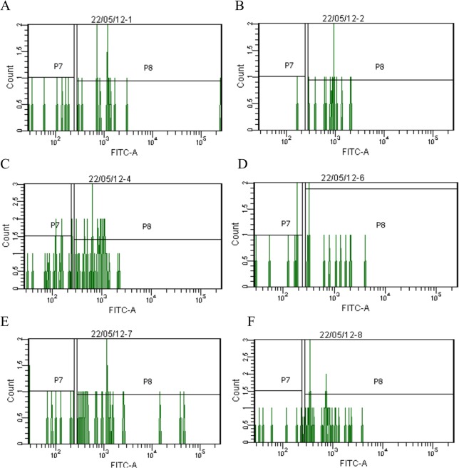
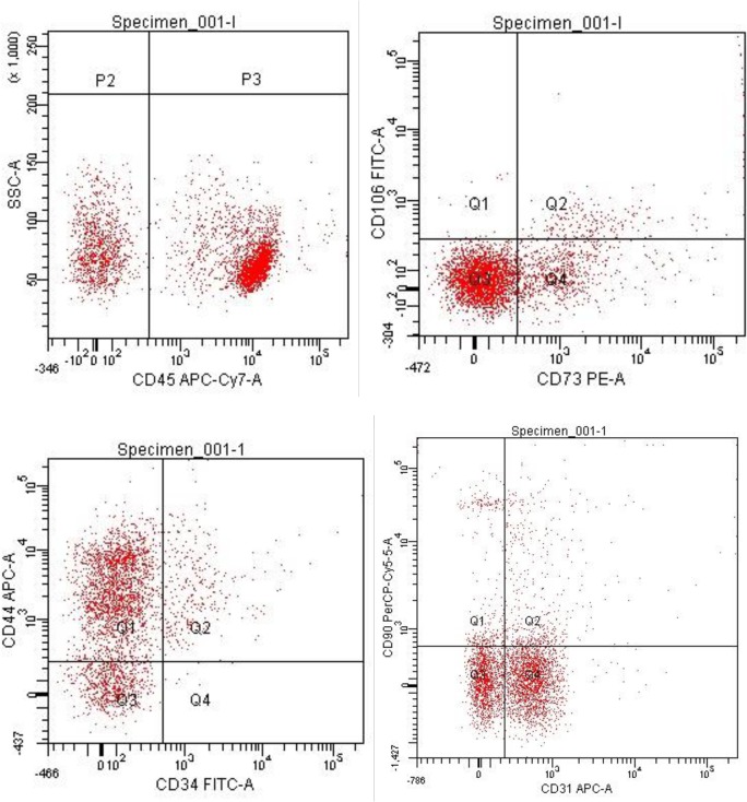
Hyaluronidases are groups of enzymes that degrade hyaluronic acid (HA). To stop enzymatic hydrolysis we modified testicular hyaluronidase (HYAL) by activated polyethylene oxide with the help of electron-beam synthesis. As a result we received pegylated hyaluronidase (pegHYAL). Spiperone is a selective D2 dopamine receptor antagonist. It was demonstrated on the model of a single bleomycin damage of alveolar epithelium that during the inflammatory phase monotherapy by pegHYAL or spiperone reduced the populations of hematopoietic stem /progenitor cells in the lung parenchyma. PegHYAL also reduced the levels of transforming growth factor (TGF)-β, interleukin (IL)-1β, tumor necrosis factor (TNF)-α in the serum and lungs, while spiperone reduced the level of the serum IL-1β. Polytherapy by spiperone and pegHYAL caused the increase of the quantity of hematopoietic stem/ progenitor cells in the lungs. Such an influx of blood cell precursors was observed on the background of considerable fall level of TGF-β and the increase level of TNF-α in the serum and lungs. These results show pegHYAL reduced the bleomycin-induced fibrosis reaction (production and accumulation of collagen) in the lung parenchyma. This effect was observed at a single and repetitive bleomycin damage of alveolar epithelium, the antifibrotic activity of pegHYAL surpassing the activity of testicular HYAL. The antifibrotic effect of pegHYAL is enhanced by an additional instillation of spiperone. Therapy by pegHYAL causes the flow of CD31‒ CD34‒ CD45‒ CD44+ CD73+ CD90+ CD106+-cells into the fibrous lungs. These cells are incapable of differentiating into fibroblast cells. Spiperone instillation separately or together with pegHYAL reduced the MSC-like cells considerably. These data enable us to assume, that pegHYAL is a new and promising instrument both for preventive and therapy of toxic pneumofibrosis. The blockage of D2 dopamine receptors with the following change of hyaluronan matrix can be considered as a new strategy in treatment of pneumofibrosis.
Conflict of interest statement
Figures
Similar articles
-
Micellar Hyaluronidase and Spiperone as a Potential Treatment for Pulmonary Fibrosis.Int J Mol Sci. 2021 May 25;22(11):5599. doi: 10.3390/ijms22115599. Int J Mol Sci. 2021. PMID: 34070506 Free PMC article.
-
Anti-inflammatory and antifibrotic effects of a combination of spiperone and immobilized hyaluronidase on partially reversible and irreversible toxic pneumofibrosis.Bull Exp Biol Med. 2013 Nov;156(1):53-8. doi: 10.1007/s10517-013-2276-0. Bull Exp Biol Med. 2013. PMID: 24319728
-
Effect of spiperone on mesenchymal multipotent stromal and hemopoietic stem cells under conditions of pulmonary fibrosis.Bull Exp Biol Med. 2014 May;157(1):132-7. doi: 10.1007/s10517-014-2508-y. Epub 2014 Jun 11. Bull Exp Biol Med. 2014. PMID: 24913578
-
Antifibrotic activity of hyaluronidase immobilized on polyethylenoxide under conditions of bleomycin-induced pneumofibrosis.Bull Exp Biol Med. 2013 Jan;154(3):388-92. doi: 10.1007/s10517-013-1957-z. Bull Exp Biol Med. 2013. PMID: 23484207
-
Gene therapy and endothelial cell targeting for cancer.Ann N Y Acad Sci. 1994 May 31;716:257-64. doi: 10.1111/j.1749-6632.1994.tb21717.x. Ann N Y Acad Sci. 1994. PMID: 7517652 Review.
Cited by
-
Dopamine receptors and organ fibrosis.Biochem Biophys Rep. 2025 Jan 6;41:101910. doi: 10.1016/j.bbrep.2024.101910. eCollection 2025 Mar. Biochem Biophys Rep. 2025. PMID: 39867679 Free PMC article. Review.
-
Peripheral-to-central immune communication at the area postrema glial-barrier following bleomycin-induced sterile lung injury in adult rats.Brain Behav Immun. 2020 Jul;87:610-633. doi: 10.1016/j.bbi.2020.02.006. Epub 2020 Feb 22. Brain Behav Immun. 2020. PMID: 32097765 Free PMC article.
-
Antifibrotic and Regenerative Effects of Treamid in Pulmonary Fibrosis.Int J Mol Sci. 2020 Nov 8;21(21):8380. doi: 10.3390/ijms21218380. Int J Mol Sci. 2020. PMID: 33171668 Free PMC article.
-
Spiperone Stimulates Regeneration in Pulmonary Endothelium Damaged by Cigarette Smoke and Lipopolysaccharide.Int J Chron Obstruct Pulmon Dis. 2021 Dec 30;16:3575-3591. doi: 10.2147/COPD.S336410. eCollection 2021. Int J Chron Obstruct Pulmon Dis. 2021. PMID: 35002229 Free PMC article.
-
Micellar Hyaluronidase and Spiperone as a Potential Treatment for Pulmonary Fibrosis.Int J Mol Sci. 2021 May 25;22(11):5599. doi: 10.3390/ijms22115599. Int J Mol Sci. 2021. PMID: 34070506 Free PMC article.
References
-
- Hubbard R, Cooper M, Antoniak M, Venn A, Khan S, Johnston I, et al. Risk of cryptogenic fibrosing alveolitis in metal workers. Lancet. 2000. February 5;355(9202): 466–467. - PubMed
-
- Baumgarter KB, Samet JM, Coultas DB, Stidley CA, Hunt WC, Colby TV, et al. Occupational and environmental risk factors for idiopathic pulmonary fibrosis: a multicenter case-control study. Am J Epidemiol. 2000. August 15;152(4): 307–315. - PubMed
-
- Weissman DN. Silicosis In Interstitial lung disease. Edited by Schwarz MI, King TE. 4th edition Ontario (Canada): BC Decker; 2003. pp. 387–401.
-
- Banks DE. Coal workers' pneumoconiosis In Interstitial lung disease. Edited by Schwarz MI, King TE. 4th edition Ontario (Canada): BC Decker; 2003. pp. 402–417.
MeSH terms
Substances
LinkOut - more resources
Full Text Sources
Other Literature Sources
Medical
Research Materials
Miscellaneous
