. 2015 Apr 25:13:131.
doi: 10.1186/s12967-015-0489-x.

Estradiol induces HOTAIR levels via GPER-mediated miR-148a inhibition in breast cancer

Affiliations

Estradiol induces HOTAIR levels via GPER-mediated miR-148a inhibition in breast cancer

Sifeng Tao et al. J Transl Med. .

Abstract

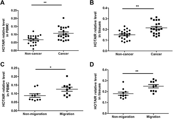
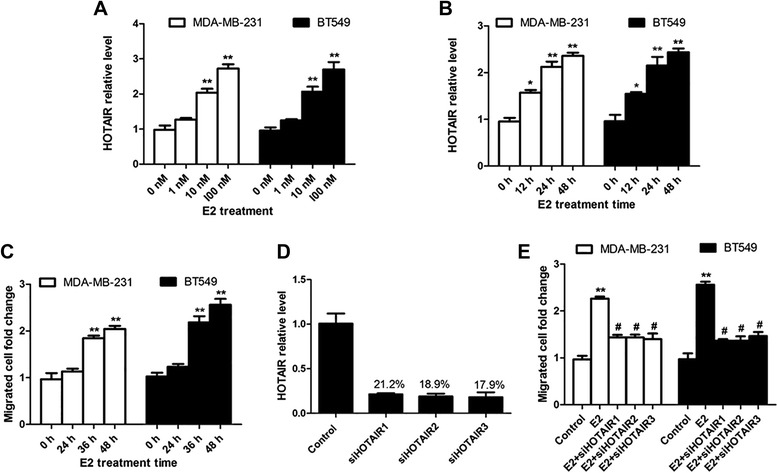
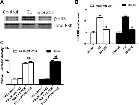
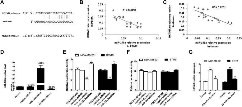
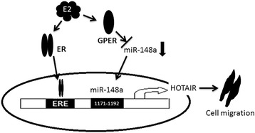
HOTAIR plays an important role in the regulation of cancer cell proliferation and cancer invasion in breast cancer. The up-regulation of HOTAIR has been reported in both estrogen receptor (ER) positive and triple-negative (TN) breast cancer. It has been reported that HOTAIR is regulated by estrogen (E2) via ERs in ER-positive breast cancer. However, it is unknown how HOTAIR is regulated in TN breast cancer. In this study, we found that HOTAIR was increased in the peripheral blood mononuclear cells and cancer tissues from breast cancer patients, and was especially higher in patients with metastatic breast cancer. In addition, we found that estrogen promoted HOTAIR through its receptor GPER and estrogen-induced breast cancer cell migration was reversed by deleting HOTAIR in TN breast cancer cells MDA-MB-231and BT549. Furthermore, we identified that E2-GPER induces the level of HOTAIR through the suppression of miR-148a. miR-148a level was negatively correlated with HOTAIR level in breast cancer patients. After the mutation of the predicted miR-148a binding sites in HOTAIR, miR-148a had no effect on HOTAIR. In conclusion, our findings offer important new insights into the ability of estrogenic GPER signaling to increase the HOTAIR level by inhibiting miR-148a in breast cancer.


Figures

Figure 1
Figure 1
HOTAIR level increased in the PBMCs and tumor tissues from the breast cancer patients. A): PBMCs were collected from 20 patients with breast cancer and 20 healthy women. Then the expression of HOTAIR was detected using real-time PCR. B): Fresh 20 human breast cancer tissues and 20 normal human breast tissues were collected and HOTAIR expression was assessed with real-time PCR. C): PBMCs were collected from 10 patients with breast cancer migration and 10 without migration. Then the expression of HOTAIR was detected using real-time PCR. D): Fresh 10 breast cancer tissues from patients with breast cancer migration and 10 without migration were collected and HOTAIR expression was assessed by using real-time PCR. *p < 0.05, **p < 0.01.
Figure 2
Figure 2
E2-induced HOTAIR increases the migration of breast cancer cells. A): MDA-MB-231 and BT549 cells were treated with the indicated concentration of E2 for 24 hours and the expression of HOTAIR was assessed using real-time PCR. B): MDA-MB-231 and BT549 cells were treated with 10 nM E2 for varying hours and the expression of HOTAIR was assessed using real-time PCR. C): MDA-MB-231 and BT549 cells were treated with 10 nM E2 for varying hours and the migrated cells were counted. D): siHOTAIR RNA sequences (si-HOTAIR1, si-HOTAIR2 and si-HOTAIR3) were transfected into MDA-MB-231 for 24 h, and then the HOTAIR level was detected by using qRT-PCR. E): MDA-MB-231 and BT549 cells were transfected with the control sequence or siHOTAIR RNA sequences before treatment with 10 Nm E2 for 36 hours and the migrated cells were counted. The results are shown as mean ± S.E. from three representative independent experiments. *p < 0.05, **P < 0.01 compared with 0 h or control. #p < 0.05 The results are shown as mean ± S.E. from three representative independent experiments. **P < 0.01 compared with control. #p < 0.05 compared with E2.
Figure 3
Figure 3
GPER mediates the promotion effect of E2 on HOTAIR expression. A): MDA-MB-231 cells were treated with 1 μM G1 with or without 100 nM G15 for 6 h. Then the expression of the p-ERK level was checked with a western blot. B): MDA-MB-231 and BT549 cells were pretreated with 100 nM G15 for 6 h before the addition of 10 nM E2 for 24 h. Then the expression of HOTAIR was determined by real-time PCR. C): The HOTAIR promoter sequence was cloned into PGL3-basic luciferase reporter plasmid, and then the control plasmid and HOTAIR promoter including plasmid were transfected into the MDA-MB-231 and BT549 cells. Then 10 nM E2 was added into these transfected cells. After E2 treatment for 24 h, the luciferase activity was detected. The results are shown as mean ± S.E. from three representative independent experiments. **P < 0.01 compared with control. #p < 0.05 compared with E2. ns p > 0.5.
Figure 4
Figure 4
miR-148a targets HOTAIR in breast cancer cells. A): HOTAIR has binding sequences for miR-148a. B): The level of miR-148 was associated with the HOTAIR mRNA level in the PBMCs from the breast cancer patients. Spearman’s correlation coefficient [rs] = r2 = 0.6492, p = 0.009. C): The level of miR-148 was associated with the HOTAIR mRNA level in the tumor tissues from the breast cancer patients. Spearman’s correlation coefficient [rs] = r2 = 0.6251, p = 0.004. D: The MDA-MB-231 cells were transfected with negative miRNA, miR-148a mimics and miR-148a for 24 h, and then the level of miR-148a was assessed using qRT-PCR. E): The luciferase activities of the MDA-MB-231 and BT549 cells were measured after co-transfection with PGL3-HOTAIR and miR-148a or its inhibitor for 24 h. The results are shown as mean ± S.E. from three representative independent experiments. **P <0.01 vs. control. F): The luciferase activity of the MDA-MB-231 and BT549 cells was measured after co-transfection with the indicated mutated PGL3-mHOTAIR constructs and miR-148a or its inhibitor for 24 h. The results are shown as mean ± S.E. from three representative independent experiments. G): The MDA-MB-231 and BT549 cells were transfected with miR-148a mimics before E2 treatment. Then the transfected cells were treated with 10 nM E2 for 24 h, and the expression of HOTAIR was checked with real-time PCR. The results are shown as mean ± S.E. from three representative independent experiments. **p <0.01 vs control, #p < 0.05 vs. E2 alone.
Figure 5
Figure 5
Model of E2-induced breast cancer cell migration via up-regulation of HOTAIR expression.

Similar articles

Cited by

References

    1. Vargo-Gogola T, Rosen JM. Modelling breast cancer: one size does not fit all. Nat Rev Cancer. 2007;7:659–72. doi: 10.1038/nrc2193. - DOI - PubMed
    1. Fidler IJ. The pathogenesis of cancer metastasis: the ‘seed and soil’ hypothesis revisited. Nat Rev Cancer. 2003;3:453–8. doi: 10.1038/nrc1098. - DOI - PubMed
    1. Espinoza-Lewis RA, Wang DZ. MicroRNAs in heart development. Cur Top Dev Biol. 2012;100:279–317. doi: 10.1016/B978-0-12-387786-4.00009-9. - DOI - PMC - PubMed
    1. Zhao G, Zhou X, Chen S, Miao H, Fan H, Wang Z, et al. Differential expression of microRNAs in decidua-derived mesenchymal stem cells from patients with pre-eclampsia. J Biomed Sci. 2014;21:81. doi: 10.1186/s12929-014-0081-3. - DOI - PMC - PubMed
    1. Ulitsky I, Bartel DP. LincRNAs: genomics, evolution, and mechanisms. Cell. 2013;154:26–46. doi: 10.1016/j.cell.2013.06.020. - DOI - PMC - PubMed