Kikuchi-Fujimoto lymphadenitis imitating metastatic melanoma on positron emission tomography: a case report
- PMID: 25928106
- PMCID: PMC4434522
- DOI: 10.1186/s12893-015-0036-y
Kikuchi-Fujimoto lymphadenitis imitating metastatic melanoma on positron emission tomography: a case report
Abstract
Background: Accurate staging is critical for decision-making for the treatment of malignant conditions. Fluoro-deoxy-glucose positron emission tomography-computed tomography (FDG PET-CT) is a highly sensitive imaging modality for the assessment of distant metastases; however false positive results are possible due to its lower specificity with detection of other hypermetabolic pathologies.
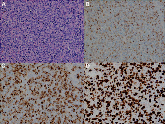
Case presentation: A patient with high-risk thigh melanoma was staged with FDG PET-CT. Four ipsilateral inguinal nodes (three superficial, one deep) demonstrated intense hypermetabolic activity. Metastatic melanoma was confirmed in the largest superficial inguinal node with ultrasound-guided fine needle aspiration. Histopathology demonstrated metastatic melanoma in one superficial node and histiocytic necrotizing lymphadenitis, also known as Kikuchi-Fujimoto disease in five deep inguinal nodes.
Conclusion: This case illustrates a false positive FDG PET-CT due to coincidental, synchronous melanoma and Kikuchi-Fujimoto disease in the same draining lymph node basin.
Figures


Similar articles
-
18F-FDG PET/CT findings in patients with Kikuchi disease.Nuklearmedizin. 2013;52(3):101-6. doi: 10.3413/Nukmed-0513-12-06. Epub 2013 Mar 27. Nuklearmedizin. 2013. PMID: 23681151
-
Lymph node uptake of 18F-fluorodeoxyglucose detected with positron emission tomography/computed tomography mimicking malignant lymphoma in a patient with Kikuchi disease.Clin Lymphoma Myeloma Leuk. 2010 Dec;10(6):477-9. doi: 10.3816/CLML.2010.n.083. Clin Lymphoma Myeloma Leuk. 2010. PMID: 21156466
-
Positron emission tomography/computed tomography hypermetabolism of Kikuchi-Fujimoto disease mimicking malignant lymphoma: a case report and literature review.J Int Med Res. 2021 Jul;49(7):3000605211032859. doi: 10.1177/03000605211032859. J Int Med Res. 2021. PMID: 34334002 Free PMC article. Review.
-
Multimodality approach of perioperative 18F-FDG PET/CT imaging, intraoperative 18F-FDG handheld gamma probe detection, and intraoperative ultrasound for tumor localization and verification of resection of all sites of hypermetabolic activity in a case of occult recurrent metastatic melanoma.World J Surg Oncol. 2008 Jan 10;6:1. doi: 10.1186/1477-7819-6-1. World J Surg Oncol. 2008. PMID: 18186915 Free PMC article.
-
[Kikuchi disease and lupus: case report, literature review and FDG PET/CT interest].J Mal Vasc. 2011 Jul;36(4):274-9. doi: 10.1016/j.jmv.2011.05.004. Epub 2011 Jul 14. J Mal Vasc. 2011. PMID: 21757306 Review. French.
Cited by
-
Kikuchi-Fujimoto disease in the regional lymph nodes with node metastasis in a patient with tongue cancer: A case report and literature review.Oncol Lett. 2017 Jul;14(1):257-263. doi: 10.3892/ol.2017.6139. Epub 2017 May 9. Oncol Lett. 2017. PMID: 28693162 Free PMC article.
-
A rare co-existence of histiocytic necrotizing lymphadenitis with metastatic papillary thyroid carcinoma and review of the literature.Diagn Pathol. 2024 Jan 13;19(1):14. doi: 10.1186/s13000-024-01441-0. Diagn Pathol. 2024. PMID: 38218846 Free PMC article. Review.
References
-
- Dosi RV, Ambaliya A, Patel JS, Bhambhani YS, Patell RD. A fatal case of Kikuchi-Fujimoto disease. J Indian Med Assoc. 2012;110(1):53–4. - PubMed
Publication types
MeSH terms
Substances
LinkOut - more resources
Full Text Sources
Other Literature Sources
Medical

