. 2015 Apr 30;10(4):e0125263.
doi: 10.1371/journal.pone.0125263. eCollection 2015.

Nanoscale Synaptic Membrane Mimetic Allows Unbiased High Throughput Screen That Targets Binding Sites for Alzheimer's-Associated Aβ Oligomers

Affiliations

Nanoscale Synaptic Membrane Mimetic Allows Unbiased High Throughput Screen That Targets Binding Sites for Alzheimer's-Associated Aβ Oligomers

Kyle C Wilcox et al. PLoS One. .

Abstract

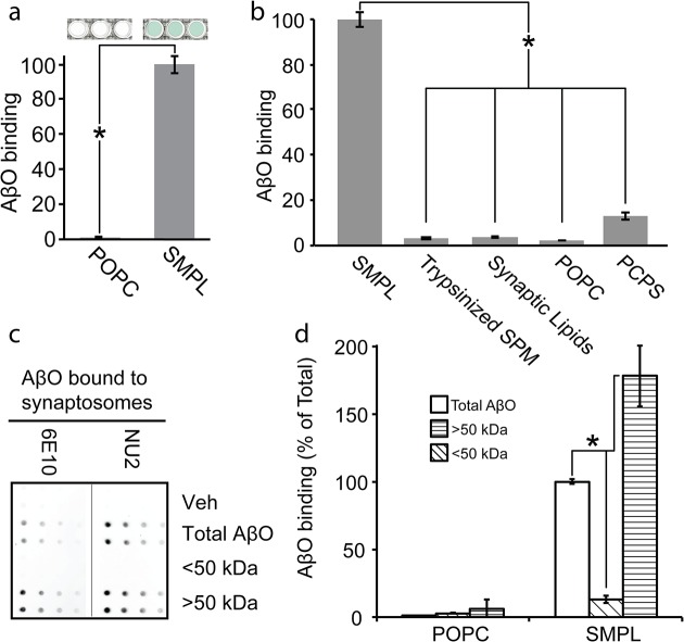
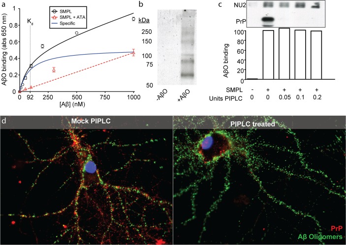
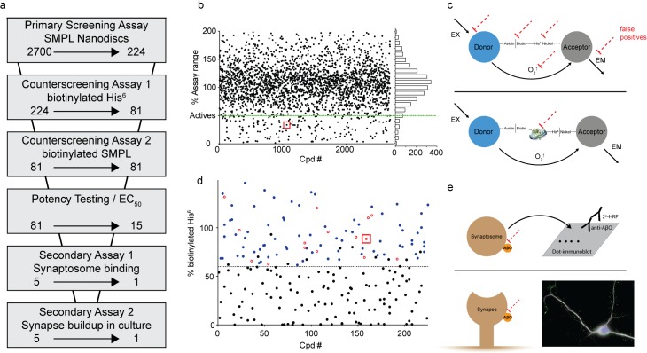
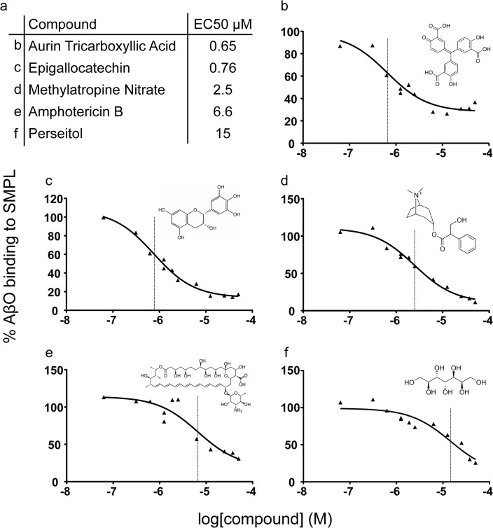
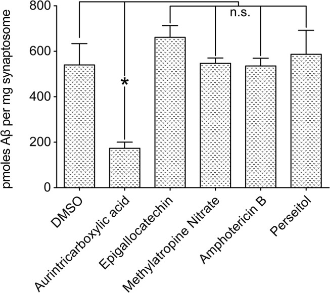
Despite their value as sources of therapeutic drug targets, membrane proteomes are largely inaccessible to high-throughput screening (HTS) tools designed for soluble proteins. An important example comprises the membrane proteins that bind amyloid β oligomers (AβOs). AβOs are neurotoxic ligands thought to instigate the synapse damage that leads to Alzheimer's dementia. At present, the identities of initial AβO binding sites are highly uncertain, largely because of extensive protein-protein interactions that occur following attachment of AβOs to surface membranes. Here, we show that AβO binding sites can be obtained in a state suitable for unbiased HTS by encapsulating the solubilized synaptic membrane proteome into nanoscale lipid bilayers (Nanodiscs). This method gives a soluble membrane protein library (SMPL)--a collection of individualized synaptic proteins in a soluble state. Proteins within SMPL Nanodiscs showed enzymatic and ligand binding activity consistent with conformational integrity. AβOs were found to bind SMPL Nanodiscs with high affinity and specificity, with binding dependent on intact synaptic membrane proteins, and selective for the higher molecular weight oligomers known to accumulate at synapses. Combining SMPL Nanodiscs with a mix-incubate-read chemiluminescence assay provided a solution-based HTS platform to discover antagonists of AβO binding. Screening a library of 2700 drug-like compounds and natural products yielded one compound that potently reduced AβO binding to SMPL Nanodiscs, synaptosomes, and synapses in nerve cell cultures. Although not a therapeutic candidate, this small molecule inhibitor of synaptic AβO binding will provide a useful experimental antagonist for future mechanistic studies of AβOs in Alzheimer's model systems. Overall, results provide proof of concept for using SMPLs in high throughput screening for AβO binding antagonists, and illustrate in general how a SMPL Nanodisc system can facilitate drug discovery for membrane protein targets.


Conflict of interest statement

Competing Interests: KCW, SGS, and WLK are affiliated with SMPL Solutions, an LLC that targets AβO binding for Alzheimer's therapeutics.

Figures

Fig 1
Fig 1. AβO receptors are highly connected.
Rat cortical synaptosomes were titrated in solution with AβOs and binding was detected by dot immunoblot (Mean +/- SD; n = 3) (a). AβO-bound synaptosomes were broken down with triton and deoxycholate to maintain AβO interactions with the binding site and detergent-lysates were immunodepleted using AβO-specific NU2 antibody. A negligible fraction of total synaptosomal protein is co-immunoprecipitated, illustrating a highly selective binding (Mean +/- SD; n = 2) (b). The co-immunoprecipitated fraction was analyzed by SDS-PAGE and silver staining to assess the number and size of proteins associated with an AβO binding complex (c).
Fig 2
Fig 2. Nanodiscs preserve synaptic protein composition and structure.
(a) Schematic of SMPL Nanodisc formation using synaptic plasma membranes. (b) Nanodiscs containing biotinylated synaptic membranes (solid curve) or POPC (dashed curve) were separated by size exclusion chromatography. Fractions eluting from the synaptic Nanodisc run were collected and analyzed by dot blot to locate biotinylated synaptic proteins (red curve; Mean +/-SD; n = 2). The population of biotinylated membrane proteins inserted into Nanodiscs was analyzed by SDS-PAGE, probing for biotin (c) or using antibodies against specific proteins related to AβO binding (d). 3H glutamate binding to SMPL Nanodiscs was assessed in the absence and presence of a 100-fold excess of cold glutamate (Mean +/- SD; n = 3; * p<0.05) (e). Enzymatic activity was assessed in synaptic plasma membranes (Syn PM) and SMPL Nanodiscs (SMPL) by probing for tyrosine phosphorylation in the absence and presence of ATP (f). Insulin receptor activity was probed using an antibody recognizing IRβpTyr1162/1163 (g).
Fig 3
Fig 3. Synaptic Nanodiscs contain an AβO binding protein that interacts selectively with high molecular weight oligomers.
A Nanodisc-based assay for AβO binding indicates the transfer of the AβO binding site into Nanodiscs (a) (n = 3; mean +/- SD; * p<0.05). Nanodiscs containing a range of detergent-solubilized precursor membranes or lipid mixtures were applied to the AβO binding assay (b; SPM—synaptic plasma membrane, Trypsinized SPM—SPM treated with trypsin prior to Nanodisc incorporation, Synaptic Lipids—lipids extracted from SPM, POPC—100% POPC lipid, PC/PS—1:1 POPC:POPS; n = 3; mean +/- SD; * p<0.05). AβOs were separated into populations smaller (diagonal fill) and larger (horizontal fill) than 50 kDa and assayed for binding to synaptosomes (c) and Nanodiscs (d). (n = 2; mean +/- SD; * p<0.05)
Fig 4
Fig 4. AβO binding is receptor-mediated but PrPC-independent in Nanodiscs and mature neurons.
Immobilized SMPL Nanodiscs were titrated with AβOs and bound AβOs were detected with NU2 oligomer-specific antibody coupled to an HRP-based colorimetric assay. Nonspecific binding was measured in the presence of excess ATA, which blocks AβO/receptor binding, and used to calculate the specific binding component (blue). n = 3; mean +/- SD. (a). To analyze Nanodisc proteins co-immunoprecipitating with AβOs, Nanodiscs containing biotinylated synaptic plasma membranes were affinity precipitated and visualized by SDS-PAGE immunoblot with biotin detection (b). To test the prediction that PrPC mediates AβO binding, immobilized Nanodiscs were split into four equivalent reactions and pre-treated with 0, 0.05, 0.1, or 0.2 units of PIPLC to remove PrPC before exposing to AβOs and probing with NU2 as in (a). PrPC removal was verified by Western blotting after the colorimetric assay was complete. NU2 bound to each immobilized Nanodisc/AβO complex is detected in the blot by anti-mouse secondary antibodies used to probe for the mouse antibody against PrPC (c). The effect of PrPC removal on AβO binding was tested using mature hippocampal cultures treated with PIPLC (d). PrPC was detected using an antibody (red) and fluorescence-conjugated AβOs were visualized directly (green).
Fig 5
Fig 5. SMPL Nanodiscs provide the basis for a high-throughput assay for AβO binding antagonists.
A schematic of the AlphaScreen assay adapted to measure AβO binding to synaptic Nanodiscs (a). Biotinylated AβOs and His-tagged MSP molecules link Nanodiscs to AlphaScreen donor and acceptor beads. The proof-of-concept assay produces high dynamic range (b). NU2 oligomer-specific antibodies were used as a drug stand-in to test the assay’s response to an applied treatment (c).
Fig 6
Fig 6. Screening strategy effectively eliminates false positives.
(a) Screening assays used to evaluate the effect of Spectrum Collection molecules on AβO binding. The arrows indicate the reduction in compounds resulting at each step. (b) Compiled data from the primary AlphaScreen assay are shown in a single graph normalized to POPC and SMPL in-plate controls. (c) Schematics of counterscreening assays designed to identify false positive compounds acting on off-target elements of the primary screening assay (dashed red lines). Assays use AlphaScreen donor and acceptor beads linked together by either biotinylated hexahistidine (top) or Nanodiscs containing biotinylated synaptic proteins (bottom). (d) Data from the biotinylated hexahistidine counterscreen. Black symbols denote compounds classified as likely false positives. Blue symbols denote compounds that were retested in dose-response format (Examples shown in Fig 7), and the compounds showing significant signal reduction at 1 μM are shown as open red circles. (e) Secondary, orthogonal assays to verify compound efficacy in preventing AβO binding include a dot immunoblot test for AβO binding to rat cortical synaptosomes (Top; shown in Fig 8) and an immunocytochemical analysis of AβO binding to cultures of rat hippocampal neurons (Bottom; shown in Fig 9 for ATA). Red squares in panels b and d identify the data points associated with ATA.
Fig 7
Fig 7. Dose response testing of selected compounds.
Of fifteen compounds surviving dose-response potency testing, five commercially-available molecules were repurchased for further analysis. (a) The names and EC50 values of each are listed with the corresponding panel identifier. (b-f) Individual dose response curves and chemical structures corresponding to compounds listed in panel a. Vertical lines mark the EC50 values in each plot.
Fig 8
Fig 8. Impact of selected compounds on synaptosome binding.
Selected compounds were repurchased and tested for an impact on AβO binding to rat cortical synaptosomes in a dot immunoblot assay. n = 3; Mean+/-SEM.
Fig 9
Fig 9. Aurin tricarboxylic acid potently reduces synaptic AβO accumulation in culture.
Aurin tricarboxylic acid was assayed at 1μM for a preventative effect on AβO accumulation at synapses in cultured rat hippocampal neurons. (a) Typical neurons after treatment with Vehicle (left), AβOs (center), or AβOs following ATA pre-treatment (right). AβOs are shown in green, neurons identified by β3 tubulin fluorescence are white, and DAPI is blue to indicate nuclei. Selected neurites are enlarged below each image to illustrate the distribution of bound AβOs. (b) Quantification of AβO intensity per neurite branch length as a percent of AβO treated neurons. *** Denotes p<0.0001.

References

    1. Cooper MA (2004) Advances in membrane receptor screening and analysis. J Mol Recognit 17: 286–315. - PubMed
    1. Dodgson K, Gedge L, Murray DC, Coldwell M (2009) A 100K well screen for a muscarinic receptor using the Epic label-free system—a reflection on the benefits of the label-free approach to screening seven-transmembrane receptors. J Recept Signal Transduct Res 29: 163–172. 10.1080/10799890903079844 - DOI - PubMed
    1. Johnson EN, Shi X, Cassaday J, Ferrer M, Strulovici B, Kunapuli P (2008) A 1,536-well [(35)S]GTPgammaS scintillation proximity binding assay for ultra-high-throughput screening of an orphan galphai-coupled GPCR. Assay Drug Dev Technol 6: 327–337. 10.1089/adt.2007.113 - DOI - PubMed
    1. Gonzalez R, Jennings LL, Knuth M, Orth AP, Klock HE, Ou W, et al. (2010) Screening the mammalian extracellular proteome for regulators of embryonic human stem cell pluripotency. Proceedings of the National Academy of Sciences of the United States of America 107: 3552–3557. 10.1073/pnas.0914019107 - DOI - PMC - PubMed
    1. Drew D, Slotboom DJ, Friso G, Reda T, Genevaux P, Rapp M, et al. (2005) A scalable, GFP-based pipeline for membrane protein overexpression screening and purification. Protein Sci 14: 2011–2017. - PMC - PubMed

Publication types

Substances