. 2015 Apr 19:12:77.
doi: 10.1186/s12974-015-0296-6.

Activated microglia/macrophage whey acidic protein (AMWAP) inhibits NFκB signaling and induces a neuroprotective phenotype in microglia

Affiliations

Activated microglia/macrophage whey acidic protein (AMWAP) inhibits NFκB signaling and induces a neuroprotective phenotype in microglia

Alexander Aslanidis et al. J Neuroinflammation. .

Abstract

Background: Microglia reactivity is a hallmark of neurodegenerative diseases. We have previously identified activated microglia/macrophage whey acidic protein (AMWAP) as a counter-regulator of pro-inflammatory response. Here, we studied its mechanisms of action with a focus on toll-like receptor (TLR) and nuclear factor κB (NFκB) signaling.

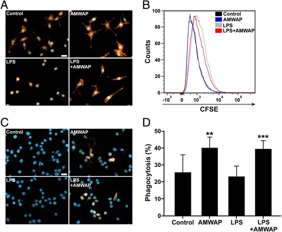
Methods: Recombinant AMWAP was produced in Escherichia coli and HEK293 EBNA cells and purified by affinity chromatography. AMWAP uptake was identified by fluorescent labeling, and pro-inflammatory microglia markers were measured by qRT-PCR after stimulation with TLR ligands. NFκB pathway proteins were assessed by immunocytochemistry, Western blot, and immunoprecipitation. A 20S proteasome activity assay was used to investigate the anti-peptidase activity of AMWAP. Microglial neurotoxicity was estimated by nitrite measurement and quantification of caspase 3/7 levels in 661W photoreceptors cultured in the presence of microglia-conditioned medium. Microglial proliferation was investigated using flow cytometry, and their phagocytosis was monitored by the uptake of 661W photoreceptor debris.

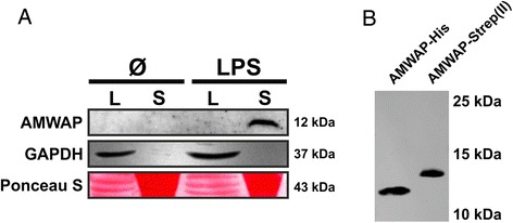
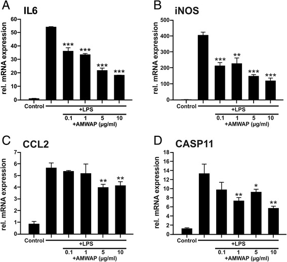
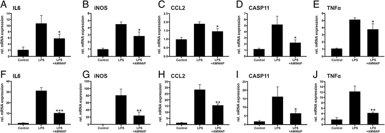
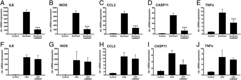
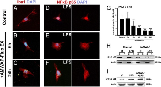
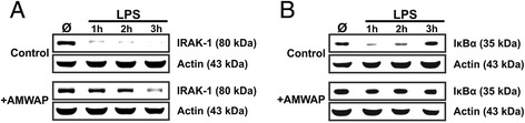
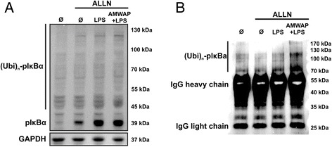
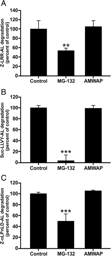
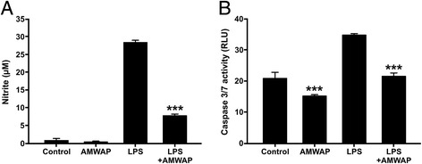
Results: AMWAP was secreted from lipopolysaccharide (LPS)-activated microglia and recombinant AMWAP reduced gene transcription of IL6, iNOS, CCL2, CASP11, and TNFα in BV-2 microglia treated with LPS as TLR4 ligand. This effect was replicated with murine embryonic stem cell-derived microglia (ESdM) and primary brain microglia. AMWAP also diminished pro-inflammatory markers in microglia activated with the TLR2 ligand zymosan but had no effects on IL6, iNOS, and CCL2 transcription in cells treated with CpG oligodeoxynucleotides as TLR9 ligand. Microglial uptake of AMWAP effectively inhibited TLR4-dependent NFκB activation by preventing IRAK-1 and IκBα proteolysis. No inhibition of IκBα phosphorylation or ubiquitination and no influence on overall 20S proteasome activity were observed. Functionally, both microglial nitric oxide (NO) secretion and 661W photoreceptor apoptosis were significantly reduced after AMWAP treatment. AMWAP promoted the filopodia formation of microglia and increased the phagocytic uptake of apoptotic 661W photoreceptor cells.

Conclusions: AMWAP is secreted from reactive microglia and acts in a paracrine fashion to counter-balance TLR2/TLR4-induced reactivity through NFκB inhibition. AMWAP also induces a neuroprotective microglial phenotype with reduced neurotoxicity and increased phagocytosis. We therefore hypothesize that anti-inflammatory whey acidic proteins could have a therapeutic potential in neurodegenerative diseases of the brain and the retina.


Figures

Figure 1
Figure 1
AMWAP is secreted from reactive BV-2 microglia. (A) BV-2 microglial cells were treated with 50 ng/ml LPS for 48 h before obtaining cellular protein lysates (L) as well as concentrated culture supernatants (S). Anti-AMWAP immunoblot analysis revealed a band of approximately 12 kDa only present in the supernatant of LPS-activated microglia. GAPDH and Ponceau S served as loading controls. (B) Anti-AMWAP immunoblot analysis of recombinantly expressed and purified AMWAP from E. coli (AMWAP-His) and HEK293 EBNA cells (AMWAP-Strep(II)) shows single bands at approximately 11 to 13 kDa.
Figure 2
Figure 2
AMWAP reduces pro-inflammatory marker gene transcription in BV-2 microglia. BV-2 cells were treated with various concentrations of recombinant AMWAP for 24 h before further stimulation with 50 ng/ml LPS for 12 h. Transcript levels of the pro-inflammatory markers IL6 (A), iNOS (B), CCL2 (C), and CASP11 (D) were determined by quantitative real-time PCR. Data show mean ± SD (n = 3/group, measured in triplicate) with *P < 0.05, **P < 0.01, ***P < 0.001 for AMWAP + LPS- vs. LPS-treated cells. PBS served as vehicle control.
Figure 3
Figure 3
AMWAP reduces TLR4-mediated pro-inflammatory gene transcription in embryonic stem cell-derived microglia and primary brain microglia. (A-E) Embryonic stem cell-derived microglia (ESdM) were treated with 10 μg/ml of recombinant AMWAP for 24 h before stimulation with 500 ng/ml LPS for further 24 h. (F-J) Primary brain microglia were treated with 10 μg/ml of recombinant AMWAP for 24 h before stimulation with 50 ng/ml LPS for further 24 h. mRNA expression of the pro-inflammatory marker transcripts IL6 (A, F), iNOS (B, G) CCL2 (C, H), CASP11 (D, I), and TNFα (E, J) was determined by quantitative real-time RT-PCR. Data show mean ± SD (n = 3/group, measured in triplicate) with *P < 0.05, **P < 0.01, ***P < 0.001 for AMWAP + LPS- vs. LPS-treated cells. PBS served as vehicle control.
Figure 4
Figure 4
AMWAP reduces TLR2-mediated pro-inflammatory gene transcription in BV-2 microglia. BV-2 cells were treated with 10 μg/ml of recombinant AMWAP for 24 h before stimulation with 50 μg/ml zymosan (A-E) or 4 μg/ml CpG oligodeoxynucleotides (F-J) for additional 24 h. The pro-inflammatory marker transcripts IL6 (A, F), iNOS (B, G), CCL2 (C, H), CASP11 (D, I), and TNFα (E, J) were measured by quantitative real-time RT-PCR. Data show mean ± SD (n = 3/group, measured in triplicate) with *P < 0.05, **P < 0.01, ***P < 0.001 for AMWAP + zymosan/CpG- vs. zymosan/CpG-treated cells. PBS served as vehicle control.
Figure 5
Figure 5
AMWAP is taken up by microglia and inhibits LPS-mediated NFκB activation. (A-C) BV-2 microglia were incubated with fluorescently labeled recombinant AMWAP (AMWAP-Fluo EX, 10 μg/ml) for 6 and 24 h. Microglia gradually incorporated AMWAP-Fluo EX into their cytosol exhibiting a perinuclear localization as shown by anti-Iba1 antibody co-staining. (D-G) AMWAP-Fluo EX pretreatment (for 1 to 24 h) time-dependently inhibited TLR4-mediated nuclear translocation of NFκB p65 after stimulation with 50 ng/ml LPS for 1 h as shown by immunocytochemistry and quantification of nuclear vs. cytosolic fluorescence intensities. PBS served as vehicle control. Data show mean ± SD (n = 9/group) with *P < 0.05, **P < 0.01, ***P < 0.001 for AMWAP-treated vs. control cells. (H) Control and AMWAP-treated BV-2 microglia were stimulated with 50 ng/ml LPS for 1 h and NFκB p65 protein amount was determined in cytosolic (c) and nuclear (n) fractions using Western blot. AMWAP-treated cells exhibited diminished LPS-triggered NFκB p65 translocation to the nucleus compared to vehicle control. (I) Control and AMWAP-treated BV-2 microglia were stimulated with 50 ng/ml LPS for 1 h and the NFκB p65 phosphorylation status (pNFκB p65) was determined in total cell lysates using Western blot. Actin served as loading control. Scale bar = 20 μm.
Figure 6
Figure 6
AMWAP prevents LPS-induced degradation of IRAK-1 and IκBα. Control and AMWAP-treated BV-2 microglia were incubated with 50 ng/ml LPS for 0, 1, 2, and 3 h, and cytosolic protein extracts were prepared. Immunoblot analysis of IRAK-1 (A) and IκBα (B) was carried out to determine the level of proteolytic degradation. In AMWAP-treated cells, both signaling molecules were protected from LPS-induced proteolysis. Resynthesized IκBα was detected after 2 and 3 h in control cells. Actin served as loading control.
Figure 7
Figure 7
AMWAP does not inhibit IκBα phosphorylation and ubiquitination. (A) Control and AMWAP-treated BV-2 microglia were preincubated with the proteasome inhibitor ALLN (100 μg/ml) for 30 min to allow for accumulation of phosphorylated IκBα before stimulation with LPS (50 ng/ml) for 30 min. Levels of phosphorylated IκBα were assessed in cytoplasmic extracts using Western blot analysis. AMWAP did not reduce LPS-induced IκBα phosphorylation. The presence of various high molecular weight bands was noticed, presumably representing polyubiquitinated, phosphorylated IκBα. (B) IκBα was immunoprecipitated from cytoplasmic samples of cells treated as in (A). Thereafter, anti-ubiquitin immunoblot revealed the presence of high molecular weight polyubiquitinated forms of phosphorylated IκBα, which were not diminished in AMWAP-positive cells. GAPDH served as loading control. (p)IκBα, (phosphorylated) inhibitor of kappa B alpha; Ubi, ubiquitin; IgG, immunoglobulin G; ALLN; N-acetyl-Leu-Leu-Norleu-al.
Figure 8
Figure 8
AMWAP does not inhibit peptidase activities of the 20S proteasome. The effect of recombinant AMWAP (20 μg/ml) on peptidase activities of the 20S proteasome was assayed using luminogenic cleavage assays with the substrates Z-LRR-aminoluciferin (trypsin-like activity) (A), Suc-LLVY-aminoluciferin (chymotrypsin-like activity) (B), and Z-nLPnLD-aminoluciferin (caspase-like activity) (C). Data show mean ± SD (n = 4/group) with *P < 0.05, **P < 0.01, ***P < 0.001 for treatment vs. control. MG-132 (1 μM), a potent 20S proteasome inhibitor, served as a positive control that reduced all three peptidase activities. PBS served as a vehicle control. MG-132, Z-Leu-Leu-Leu-al.
Figure 9
Figure 9
AMWAP reduces pro-inflammatory microglial nitric oxide production and neurotoxicity on photoreceptor cells. (A) Production of nitric oxide (NO) as determined by detection of nitrite from BV-2 microglial cells pre-treated with 10 μg/ml AMWAP or vehicle for 24 h before stimulation with 50 ng/ml LPS for further 24 h. Data show mean ± SD (n = 6/group) with *P < 0.05, **P < 0.01, ***P < 0.001 for AMWAP + LPS vs. LPS-treated cells. (B) 661W photoreceptor cells were incubated with conditioned media from vehicle-, 10 μg/ml AMWAP-, 50 ng/ml LPS-, and 50 ng/ml LPS + 10 μg/ml AMWAP-treated BV-2 microglia for 48 h and apoptosis-related caspase 3/7 activity was determined. Data show mean ± SD (n = 5/group) with *P < 0.05, **P < 0.01, ***P < 0.001 for AMWAP-treated cells vs. control and AMWAP + LPS vs. LPS-treated cells, respectively. PBS served as a vehicle control. NO, nitric oxide; RLU, relative luciferase units.
Figure 10
Figure 10
AMWAP promotes microglial filopodia formation and phagocytosis. (A) Representative images of Phalloidin-TRITC labeled BV-2 microglial cells pre-treated with 10 μg/ml AMWAP or vehicle for 24 h in the absence or presence of 50 ng/ml LPS for 24 h. (B) CFSE-proliferation assay of BV-2 microglia treated with 10 μg/ml AMWAP or vehicle for 24 h in the absence or presence of 50 ng/ml LPS for 24 h. The proliferation rate was assessed 24 h after LPS treatment using flow cytometry and a representative graph out of three repetitions is shown. (C, D) Phagocytosis of CM-DiI-stained apoptotic 661W photoreceptor debris by BV-2 microglia was monitored and quantified after 6-h feeding time of AMWAP- vs. vehicle-treated cells in the absence or presence of 50 ng/ml LPS for 24 h. Data show mean ± SD (n = 9/group) with *P < 0.05, **P < 0.01, ***P < 0.001 for AMWAP-treated cells vs. control and AMWAP + LPS vs. LPS-treated cells, respectively. PBS served as a vehicle control. CFSE, carboxyfluorescein diacetate succinimidyl ester. Scale bar = 20 μm.

References

    1. Hanisch UK, Kettenmann H. Microglia: active sensor and versatile effector cells in the normal and pathologic brain. Nat Neurosci. 2007;10:1387–94. doi: 10.1038/nn1997. - DOI - PubMed
    1. Streit WJ. Microglia as neuroprotective, immunocompetent cells of the CNS. Glia. 2002;40:133–9. doi: 10.1002/glia.10154. - DOI - PubMed
    1. Giulian D, Li J, Bartel S, Broker J, Li X, Kirkpatrick JB. Cell surface morphology identifies microglia as a distinct class of mononuclear phagocyte. J Neurosci. 1995;15:7712–26. - PMC - PubMed
    1. Davalos D, Grutzendler J, Yang G, Kim JV, Zuo Y, Jung S, et al. ATP mediates rapid microglial response to local brain injury in vivo. Nat Neurosci. 2005;8:752–8. doi: 10.1038/nn1472. - DOI - PubMed
    1. Nimmerjahn A, Kirchhoff F, Helmchen F. Resting microglial cells are highly dynamic surveillants of brain parenchyma in vivo. Science. 2005;308:1314–8. doi: 10.1126/science.1110647. - DOI - PubMed

Publication types

MeSH terms

LinkOut - more resources