. 2015 Apr 30;4(5):e001889.
doi: 10.1161/JAHA.115.001889.

Impaired development of left anterior heart field by ectopic retinoic acid causes transposition of the great arteries

Affiliations

Impaired development of left anterior heart field by ectopic retinoic acid causes transposition of the great arteries

Mayu Narematsu et al. J Am Heart Assoc. .

Abstract

Background: Transposition of the great arteries is one of the most commonly diagnosed conotruncal heart defects at birth, but its etiology is largely unknown. The anterior heart field (AHF) that resides in the anterior pharyngeal arches contributes to conotruncal development, during which heart progenitors that originated from the left and right AHF migrate to form distinct conotruncal regions. The aim of this study is to identify abnormal AHF development that causes the morphology of transposition of the great arteries.

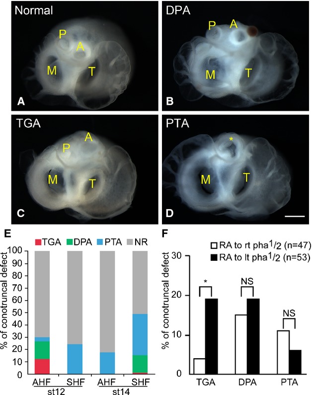
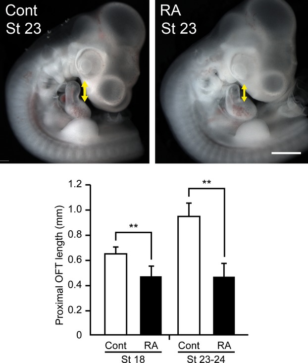
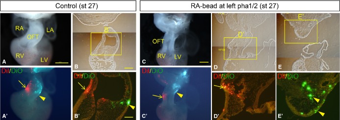
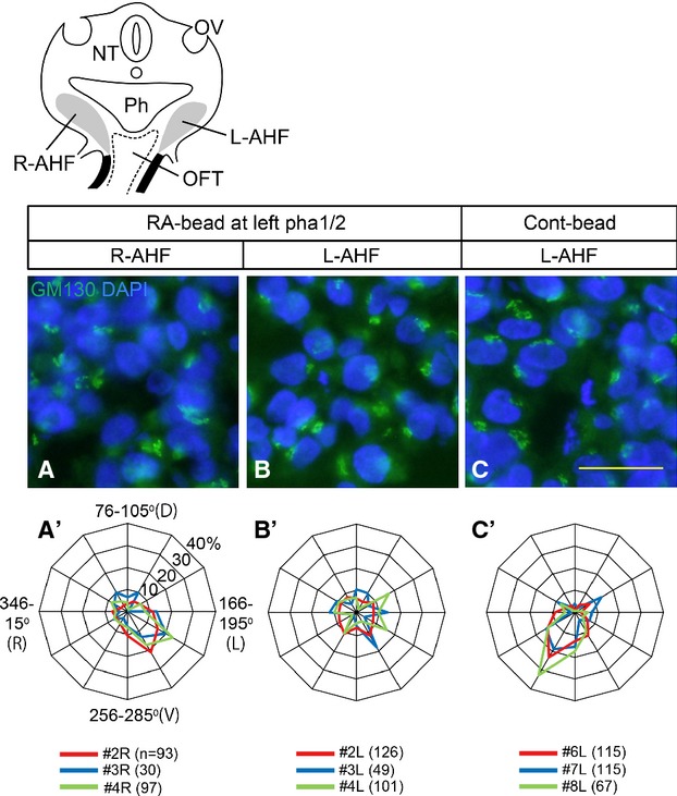
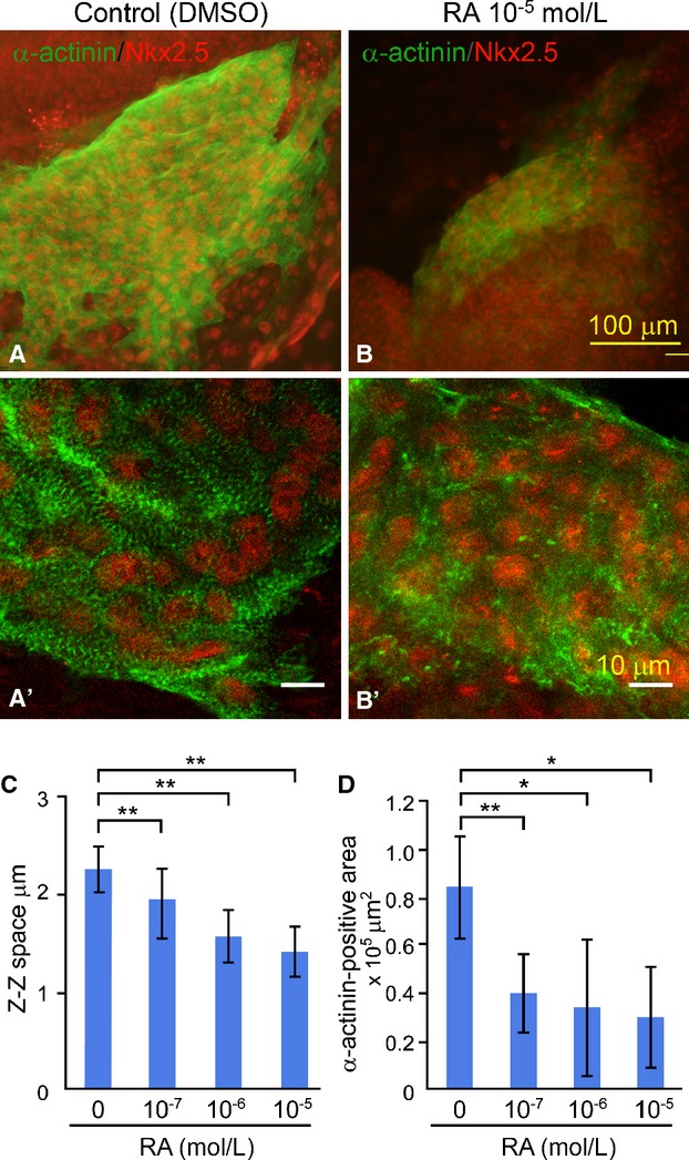
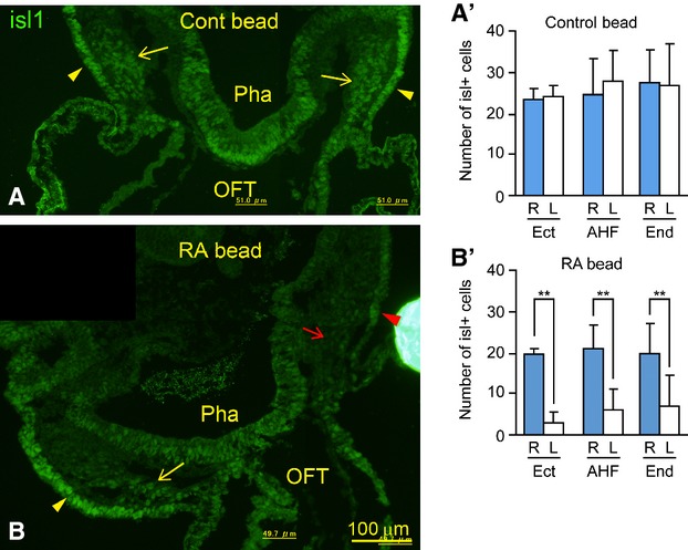
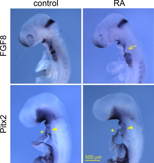
Methods and results: We placed a retinoic acid-soaked bead on the left or the right or on both sides of the AHF of stage 12 to 14 chick embryos and examined the conotruncal heart defect at stage 34. Transposition of the great arteries was diagnosed at high incidence in embryos for which a retinoic acid-soaked bead had been placed in the left AHF at stage 12. Fluorescent dye tracing showed that AHF exposed to retinoic acid failed to contribute to conotruncus development. FGF8 and Isl1 expression were downregulated in retinoic acid-exposed AHF, and differentiation and expansion of cardiomyocytes were suppressed in cultured AHF in medium supplemented with retinoic acid.

Conclusions: The left AHF at the early looped heart stage, corresponding to Carnegie stages 10 to 11 (28 to 29 days after fertilization) in human embryos, is the region of the impediment that causes the morphology of transposition of the great arteries.

Keywords: Isl1; congenital; heart defects; morphogenesis; transposition of great vessel.


Figures

Figure 1
Figure 1
RA-soaked bead placed on left AHF caused TGA morphology. A through E, An RA-soaked (0.5 mg/mL) or control (dimethyl sulfoxide–soaked) bead was placed on AHF (pha1/2) or SHF (visceral mesoderm dorsal to the heart outflow tract) at stage 12 or 14. Embryos were reincubated and sacrificed at stage 34 (embryonic day 8), and conotruncal heart defects were inspected. Hearts viewed from the atrial side show a normal position of the great arteries (A), DPA (B), TGA (C), and PTA (D). When AHF was exposed to an RA-soaked bead at stage 12, TGA was diagnosed in 12% (4 of 34), DPA was diagnosed in 15% (5 of 34), and PTA was diagnosed in 3% (1 of 34). Neither TGA nor DPA was diagnosed if SHF was exposed to an RA-soaked bead at stage 12 (0 of 17) or AHF was exposed at stage 14 (0 of 17). RA-soaked beads placed on the left and right SHF at stage 14 induced PTA (33%, 9 of 27), DPA (15%, 4 of 27), and TGA (4%, 1 of 27) (E). The incidence of TGA/DPA was significantly high in the group of RA-exposed AHF at stage 12 versus RA-exposed SHF at stage 14 (P<0.05, Fisher’s exact test). The incidence of TGA was significantly high when the left AHF was exposed to an RA-soaked bead (F; *P<0.05, Fisher’s exact test). Bar, 500 μm. A indicates aorta; AHF, anterior heart field; DMSO, dimethyl sulfoxide; DPA, dextroposed aorta; lt, left; M, mitral valve; NR, normal; NS, no significant difference; P, pulmonary trunk; pha1/2, pharyngeal arches 1 and 2; PTA, persistent truncus arteriosus; RA, retinoic acid; rt, right; SHF, secondary heart field; st, stage; T, tricuspid valve; TGA, transposition of the great arteries.
Figure 2
Figure 2
OFT is truncated in embryos treated with an RA-soaked bead. Stage 12 embryos in which an RA-soaked bead (0.5 mg/mL) or a control bead was placed at both left and right pharyngeal arches 1/2 were incubated, and the length of the proximal OFT was measured at stage 18 (5 control embryos, 8 RA-exposed embryos) and stages 23 to 24 (6 control embryos, 7 RA-exposed embryos). The length of the proximal OFT in embryos implanted with an RA-soaked bead was significantly shorter than that in control. Note that the length was measured between the base and the top of the proximal OFT (double arrow). **P<0.01 (Mann–Whitney U test). Bar, 1 mm. Cont indicates dimethyl sulfoxide–soaked bead; OFT, outflow tract; RA, retinoic-acid-soaked bead; St, stage.
Figure 3
Figure 3
RA-exposed left AHF cells failed to migrate to the OFT. An RA-soaked bead was placed on the left pha1/2, of which the mesenchymal core (AHF) had been labeled with DiO (green). The right AHF was marked with DiI (red) and treated with a control bead. Embryos were observed at stage 27 (embryonic day 5) under a fluorescent stereoscopic microscope. In control embryos, DiO-labeled cells from the left AHF migrated to the left side of the conotruncus (arrowhead in A’ and B’) and DiI-labeled cells from the right AHF migrated to the right side (arrow in A’ and B’). In embryos in which an RA-soaked bead (0.5 mg/mL) was placed on the left pha1/2 (AHF), DiI-labeled cells from the right AHF cells migrated to the right proximal OFT (arrow C’ and D’), whereas DiO-labeled left AHF cells remained in the distal OFT and pharyngeal region (18 of 21; arrowheads in C’ and E’). Bars, 500 μm (A, A’ and C, C’); 250 μm (B, D, and E); 100 μm (B’, D’, and E’). AHF indicates anterior heart field; DiI, 1,1′-dioctadecyl-3,3,3′,3′-tetramethyl-indocarbocyanine perchlorate; DiO, 3,3′-dioctadecyloxacarbocyanine perchlorate; LA, left atrium; LV, left ventricle; OFT, outflow tract; pha1/2, pharyngeal arches 1 and 2; RA, retinoic acid; RV, right ventricle; st, stage.
Figure 4
Figure 4
Cellular polarity was affected in RA-treated left AHF. Cellular polarity of AHF cells was examined. An RA-soaked bead or control bead was placed on left pha1/2 at stage 12 and reincubated for 6 hours, and the resulting embryos were stained with anti-GM130 (marker for Golgi). A clockwise angle between the vector from the center of nucleus to Golgi and right–left axis (0° to 180°) was measured in individual AHF cells. The percentage of cell number in each angle range was plotted on radar graphs, which showed a similar distribution pattern of data obtained from 3 embryos in each graph (A’, R-AHF, P=0.91; B’, RA-treated L-AHF, P=0.34; C, control L-AHF, P=0.051, χ2 test). In the right AHF and control bead placed left AHF, the vector angle was frequently distributed in 166° to 255° (A’, P<0.01) and 256° to 345° (C’, P<0.01), respectively, whereas polarity of the vector angle was not significant in RA-treated left AHF (B’, P=0.13, χ2 test). Schematic drawing shows a cross-section of the second pharyngeal arch region containing the AHF. Bar, 25 μm. AHF indicates anterior heart field; Cont, dimethyl sulfoxide–soaked bead; L, left; n, number of cells examined; NT, neural tube; OFT, outflow tract; OV, otic vesicle; Ph, pharynx; pha1/2, pharyngeal arches 1 and 2; R, right; RA, retinoic acid.
Figure 5
Figure 5
RA inhibits cardiomyocyte development in cultured left AHF. Left pharyngeal arch 2 was prepared from stage 12 embryos and cultured in medium supplemented with or without RA for 30 hours. Cultures were doubly stained with anti-sarcomeric α-actinin and anti-Nkx2.5 antibodies. In control cultures, terminally differentiated cardiomyocytes consisting of well-developed sarcomeric actinin-positive Z-bands were observed (A and A’), whereas in RA-treated cultures, there were immature cardiomyocytes consisting of premature Z-bands (bead-like deposition of sarcomeric α-actinin) (B and B’). The space between Z-bands (Z-bodies) in RA-treated explants (10−7 mol/L [4 explants, 1.92±0.33 μm], 10−6 mol/L [6 explants, 1.55±0.27], 10−5 mol/L [4 explants, 1.4±0.26]) was significantly narrow in comparison with that in the control (9 explants, 2.25±0.22) (C). The sarcomeric α-actinin/Nkx2.5-positive area in control explants (n=8, 0.82±0.25 μm2×105) (A and D) was significantly larger than that in RA-treated explants (10−7 mol/L [n=8, 0.4±0.17 μm2], 10−6 mol/L [n=8, 0.34±0.29], 10−5 mol/L [n=4, 0.30±0.21]) (B and D). A and B, conventional fluorescent microscopic images; A’ and B’, confocal microscopic images; *P<0.05; **P<0.01 (Mann–Whitney U test after Bonferroni correction). AHF indicates anterior heart field; DMSO, dimethyl sulfoxide; RA, retinoic acid.
Figure 6
Figure 6
Isl1-positive cells were decreased in the left pharyngeal region treated with an RA-soaked bead. An RA-soaked (0.5 mg/mL) or dimethyl sulfoxide–soaked (control) bead was placed on the ectoderm of left pharyngeal arches 1/2 of stage 12 embryo. After 6 hours of reincubation, stage 14 embryos were sacrificed and stained with anti-Isl1 antibody. In control embryos, there was no significant difference in the number of Isl1-positive cells between left and right pharyngeal regions (A and A’; 5 sections from 4 embryos). In contrast, the number Isl1-positive cells in left pharyngeal ectoderm (red arrowhead), AHF (red arrow), or endoderm was significantly decreased in embryos treated with an RA-soaked bead (B and B’; 9 sections from 5 embryos). Note that (A and B) were constructed from 2 photographs obtained with a ×20 objective lens (photographs obtained at ×10 are shown in Figure S5). **P<0.01 (Wilcoxon signed rank test). AHF indicates anterior heart field; Cont, control; Ect, pharyngeal ectoderm; End, pharyngeal endoderm; L, left; OFT, heart outflow tract; Pha, pharynx; R, right; RA, retinoic acid.
Figure 7
Figure 7
Fgf8 was downregulated in RA-treated AHF. Stage 12 embryos were treated with RA (or control) bead at left pharyngeal arches 1 and 2, reincubated, and sacrificed at stage 14 to 15, and the expression of Fgf8 and Pitx2 were examined. In control embryos, Fgf8 was expressed in left and right pharyngeal regions, in which AHF/SHF resides. In RA-treated embryos, the expression of Fgf8 was downregulated in the left AHF, especially in pharyngeal arch 2 (arrow), on which the RA-soaked bead had been placed. Pitx2 was expressed in left splanchnic mesoderm (arrowheads) and left side of the OFT (*) in both control and RA-exposed embryos. There was no apparent difference in the expression of Pitx2 between control and RA-treated pharyngeal region/OFT. Note that several embryos are shown in Figure S8. AHF indicates anterior heart field; OFT, heart outflow tract; RA, retinoic acid; SHF, secondary heart field.

Similar articles

Cited by

References

    1. Hoffman JI, Kaplan S. The incidence of congenital heart disease. J Am Coll Cardiol. 2002;39:1890–1900. - PubMed
    1. Ferencz C, Brenner JI, Loffredo C, Kappetein AP, Wilson PD. Transposition of the great arteries: etiologic distinctions of outflow tract defects in a case control study of risk factors. In: Clark EB, Markwald RR, Takao A, editors. Developmental Mechanisms of Heart Disease. New York, NY: Futura Publishing; 1995. pp. 639–653.
    1. Unolt M, Putotto C, Silvestri LM, Marino D, Scarabotti A, Valerio M, Caiaro A, Versacci P, Marino B. Transposition of great arteries: new insights into the pathogenesis. Front Pediatr. 2013;1:11. - PMC - PubMed
    1. Bostrom MP, Hutchins GM. Arrested rotation of the outflow tract may explain double-outlet right ventricle. Circulation. 1988;77:1258–1265. - PubMed
    1. Lomonico MP, Bostrom MP, Moore GW, Hutchins GM. Arrested rotation of the outflow tract may explain tetralogy of Fallot and transposition of the great arteries. Pediatr Pathol. 1988;8:267–281. - PubMed

Publication types

MeSH terms

Substances

LinkOut - more resources