Painful lumbosacral plexopathy: a case report
- PMID: 25929915
- PMCID: PMC4603039
- DOI: 10.1097/MD.0000000000000766
Painful lumbosacral plexopathy: a case report
Abstract
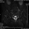
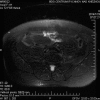
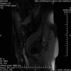
Patients frequently suffer from lumbosacral plexus disorder. When conducting a neurological examination, it is essential to assess the extent of muscle paresis, sensory disorder distribution, pain occurrence, and blocked spine. An electromyography (EMG) can confirm axonal lesions and their severity and extent, root affliction (including dorsal branches), and disorders of motor and sensory fiber conduction. Imaging examination, particularly gadolinium magnetic resonance imaging (MRI) examination, ensues. Cerebrospinal fluid examination is of diagnostic importance with radiculopathy, neuroinfections, and for evidence of immunoglobulin synthesis. Differential diagnostics of lumbosacral plexopathy (LSP) include metabolic, oncological, inflammatory, ischemic, and autoimmune disorders.In the presented case study, a 64-year-old man developed an acute onset of painful LSP with a specific EMG finding, MRI showing evidence of plexus affliction but not in the proximal part of the roots. Painful plexopathy presented itself with severe muscle paresis in the femoral nerve and the obturator nerve innervation areas, and gradual remission occurred after 3 months. Autoimmune origin of painful LSP is presumed.We describe a rare case of patient with painful lumbar plexopathy, with EMG findings of axonal type, we suppose of autoimmune etiology.
Conflict of interest statement
The authors have no funding and conflicts of interest to disclose.
Figures


References
-
- Planner AC, Donaghy M, Moore NR. Causes of lumbosacral plexopathy. Clin Radiol 2006; 61:987–995. - PubMed
-
- Barr K. Electrodiagnosis of lumbar radiculopathy. Phys Med Rehabil Clin Am 2013; 24:79–91. - PubMed
-
- Pourmand R. Immune-Mediated Neuromuscular Diseases. Basel: Karger; 2009.
-
- Laughlin RS, Dyck PJB. Electrodiagnostic testing in lumbosacral plexopathies. Phys Med Rehab Clin N Am 2013; 24:93–105. - PubMed
-
- Wilbourn AJ. Plexopathies. Neurol Clin 2007; 25:139–171. - PubMed
Publication types
MeSH terms
Substances
LinkOut - more resources
Full Text Sources
Medical

