

Near-ultraviolet laser diodes for brilliant ultraviolet fluorophore excitation
- PMID: 25930008
- PMCID: PMC8335900
- DOI: 10.1002/cyto.a.22686
Near-ultraviolet laser diodes for brilliant ultraviolet fluorophore excitation
Abstract
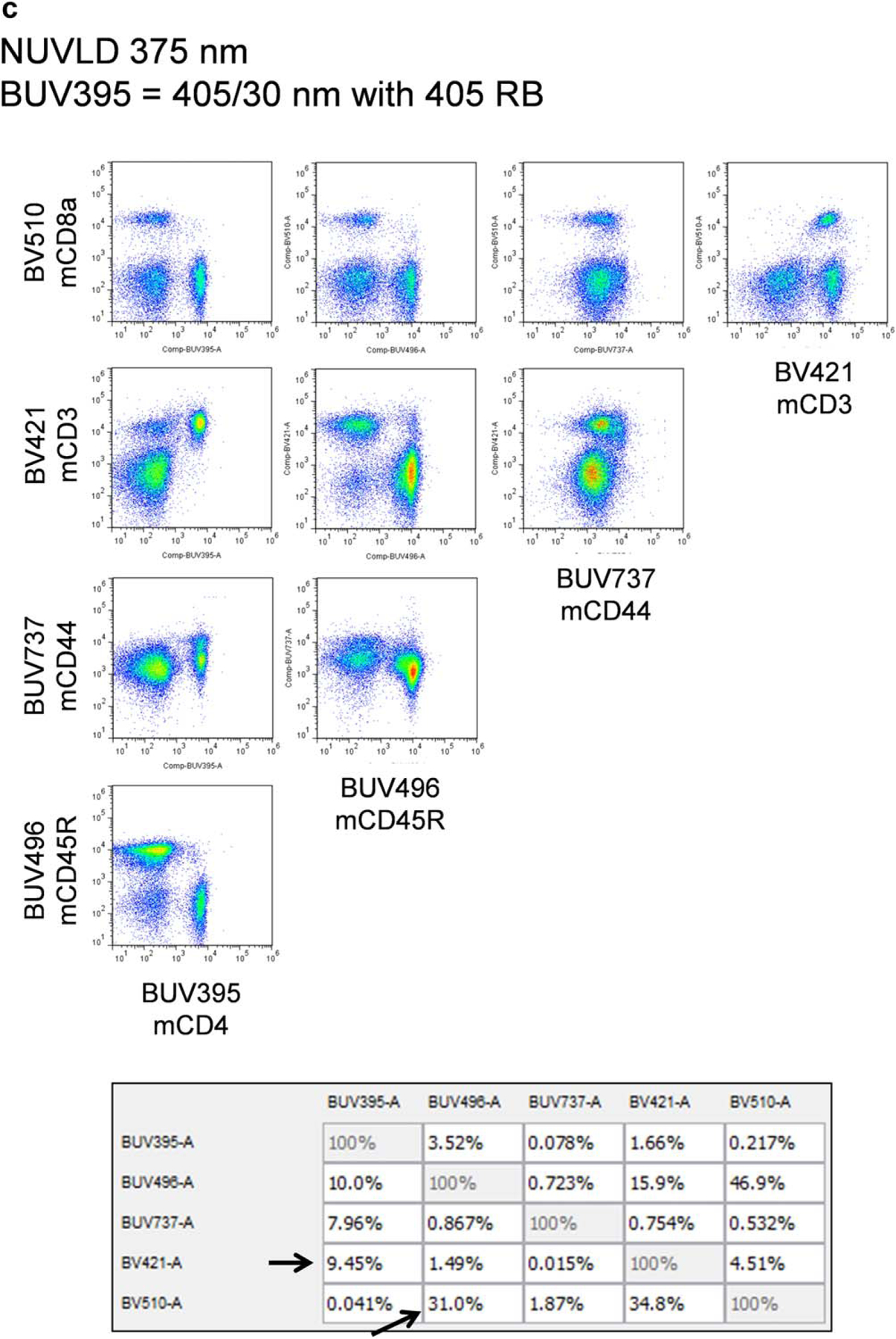
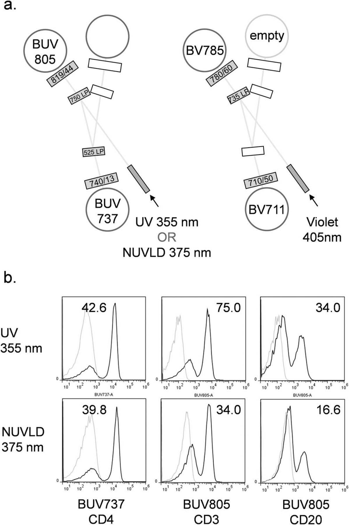
Although multiple lasers are now standard equipment on most modern flow cytometers, ultraviolet (UV) lasers (325-365 nm) remain an uncommon excitation source for cytometry. Nd:YVO4 frequency-tripled diode pumped solid-state lasers emitting at 355 nm are now the primary means of providing UV excitation on multilaser flow cytometers. Although a number of UV excited fluorochromes are available for flow cytometry, the cost of solid-state UV lasers remains prohibitively high, limiting their use to all but the most sophisticated multilaser instruments. The recent introduction of the brilliant ultraviolet (BUV) series of fluorochromes for cell surface marker detection and their importance in increasing the number of simultaneous parameters for high-dimensional analysis has increased the urgency of including UV sources in cytometer designs; however, these lasers remain expensive. Near-UV laser diodes (NUVLDs), a direct diode laser source emitting in the 370-380 nm range, have been previously validated for flow cytometric analysis of most UV-excited probes, including quantum nanocrystals, the Hoechst dyes, and 4',6-diamidino-2-phenylindole. However, they remain a little-used laser source for cytometry, despite their significantly lower cost. In this study, the ability of NUVLDs to excite the BUV dyes was assessed, along with their compatibility with simultaneous brilliant violet (BV) labeling. A NUVLD emitting at 375 nm was found to excite most of the available BUV dyes at least as well as a UV 355 nm source. This slightly longer wavelength did produce some unwanted excitation of BV dyes, but at sufficiently low levels to require minimal additional compensation. NUVLDs are compact, relatively inexpensive lasers that have higher power levels than the newest generation of small 355 nm lasers. They can, therefore, make a useful, cost-effective substitute for traditional UV lasers in multicolor analysis involving the BUV and BV dyes.
Keywords: NUVLD; brilliant ultraviolet dye; flow cytometer; near-ultraviolet laser diode; ultraviolet laser.
Published 2015 Wiley Periodicals Inc. on behalf of ISAC.
Conflict of interest statement
The author has no financial interests in any company or technology cited in this study.
Figures


Similar articles
-
Ultraviolet 320 nm laser excitation for flow cytometry.Cytometry A. 2017 Apr;91(4):314-325. doi: 10.1002/cyto.a.23066. Epub 2017 Feb 27. Cytometry A. 2017. PMID: 28240810 Free PMC article.
-
Deep ultraviolet lasers for flow cytometry.Cytometry A. 2019 Feb;95(2):227-233. doi: 10.1002/cyto.a.23640. Epub 2018 Nov 13. Cytometry A. 2019. PMID: 30423208
-
Analysis of UV-excited fluorochromes by flow cytometry using near-ultraviolet laser diodes.Cytometry A. 2004 Sep;61(1):9-17. doi: 10.1002/cyto.a.20032. Cytometry A. 2004. PMID: 15351984
-
Laser Sources for Traditional and Spectral Flow Cytometry.Methods Mol Biol. 2024;2779:33-68. doi: 10.1007/978-1-0716-3738-8_3. Methods Mol Biol. 2024. PMID: 38526781 Review.
-
Capillary electrophoresis hyphenated with UV-native-laser induced fluorescence detection (CE/UV-native-LIF).Electrophoresis. 2017 Jan;38(1):135-149. doi: 10.1002/elps.201600248. Epub 2016 Sep 5. Electrophoresis. 2017. PMID: 27445082 Review.
Cited by
-
Detection of Rare Objects by Flow Cytometry: Imaging, Cell Sorting, and Deep Learning Approaches.Int J Mol Sci. 2020 Mar 27;21(7):2323. doi: 10.3390/ijms21072323. Int J Mol Sci. 2020. PMID: 32230871 Free PMC article. Review.
-
OMIP-095: 40-Color spectral flow cytometry delineates all major leukocyte populations in murine lymphoid tissues.Cytometry A. 2023 Nov;103(11):839-850. doi: 10.1002/cyto.a.24788. Epub 2023 Sep 28. Cytometry A. 2023. PMID: 37768325 Free PMC article.
-
Fluorescent Proteins for Flow Cytometry.Curr Protoc Cytom. 2017 Apr 3;80:9.12.1-9.12.20. doi: 10.1002/cpcy.17. Curr Protoc Cytom. 2017. PMID: 28369764 Free PMC article.
-
Bioconjugatable, PEGylated Hydroporphyrins for Photochemistry and Photomedicine. Narrow-Band, Near-Infrared-Emitting Bacteriochlorins.New J Chem. 2016 Sep 1;40(9):7750-7767. doi: 10.1039/C6NJ01155A. Epub 2016 Jul 22. New J Chem. 2016. PMID: 28133433 Free PMC article.
-
Ultraviolet 320 nm laser excitation for flow cytometry.Cytometry A. 2017 Apr;91(4):314-325. doi: 10.1002/cyto.a.23066. Epub 2017 Feb 27. Cytometry A. 2017. PMID: 28240810 Free PMC article.
References
-
- Shapiro HM, Telford WG. Lasers for flow cytometry. In: Robinson JP, Darzynkiewicz Z, Dobrucki J, Hoffman RA, Nolan JP, Orfao A, Rabinovitch PS, editors. Current Protocols in Cytometry. New York: Wiley; 2009. p 1.9.
-
- Shapiro HM. Trends and developments in flow cytometry instrumentation in clinical flow cytometry. Ann N Y Acad Sci 1993; 677:155–166. - PubMed
-
- Doornbos RMP, De Grooth BG, Kraan YM, Van Der Poel CJ, Greve J. Visible diode lasers can be used for flowcytometric immunofluorescence and DNA analysis. Cytometry 1994; 15:267–271. - PubMed
-
- Shapiro HM, Perlmutter NG. Violet laser diodes as light sources for cytometry. Cytometry 2001; 44:133–136. - PubMed
-
- Telford WG, Hawley TS, Hawley RG. Analysis of violet-excited fluorochromes by flow cytometry using a violet laser diode. Cytometry Part A 2003; 54A:48–55. - PubMed
MeSH terms
Substances
Grants and funding
LinkOut - more resources
Full Text Sources
