TLR2 and TLR9 contribute to alcohol-mediated liver injury through induction of CXCL1 and neutrophil infiltration
- PMID: 25930080
- PMCID: PMC4491507
- DOI: 10.1152/ajpgi.00031.2015
TLR2 and TLR9 contribute to alcohol-mediated liver injury through induction of CXCL1 and neutrophil infiltration
Abstract
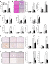
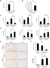
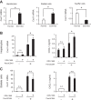
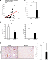
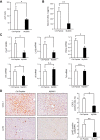
Although previous studies reported the involvement of the TLR4-TRIF pathway in alcohol-induced liver injury, the role of TLR2 and TLR9 signaling in alcohol-mediated neutrophil infiltration and liver injury has not been elucidated. Since alcohol binge drinking is recognized to induce more severe form of alcohol liver disease, we used a chronic-binge ethanol-feeding model as a mouse model for early stage of alcoholic hepatitis. Whereas a chronic-binge ethanol feeding induced alcohol-mediated liver injury in wild-type mice, TLR2- and TLR9-deficient mice showed reduced liver injury. Induction of neutrophil-recruiting chemokines, including Cxcl1, Cxcl2, and Cxcl5, and hepatic neutrophil infiltration were increased in wild-type mice, but not in TLR2- and TLR9-deficient mice. In vivo depletion of Kupffer cells (KCs) by liposomal clodronate reduced liver injury and the expression of Il1b, but not Cxcl1, Cxcl2, and Cxcl5, suggesting that KCs are partly associated with liver injury, but not neutrophil recruitment, in a chronic-binge ethanol-feeding model. Notably, hepatocytes and hepatic stellate cells (HSCs) produce high amounts of CXCL1 in ethanol-treated mice. The treatment with TLR2 and TLR9 ligands synergistically upregulated CXCL1 expression in hepatocytes. Moreover, the inhibitors for CXCR2, a receptor for CXCL1, and MyD88 suppressed neutrophil infiltration and liver injury induced by chronic-binge ethanol treatment. Consistent with the above findings, hepatic CXCL1 expression was highly upregulated in patients with alcoholic hepatitis. In a chronic-binge ethanol-feeding model, the TLR2 and TLR9-dependent MyD88-dependent pathway mediates CXCL1 production in hepatocytes and HSCs; the CXCL1 then promotes neutrophil infiltration into the liver via CXCR2, resulting in the development of alcohol-mediated liver injury.
Keywords: AH; ALD; MyD88; binge ethanol feeding; chemokine.
Copyright © 2015 the American Physiological Society.
Figures

Similar articles
-
The critical role of toll-like receptor (TLR) 4 in alcoholic liver disease is independent of the common TLR adapter MyD88.Hepatology. 2008 Oct;48(4):1224-31. doi: 10.1002/hep.22470. Hepatology. 2008. PMID: 18792393 Free PMC article.
-
Deletion of C-C Motif Chemokine Ligand 5 Worsens Invariant Natural Killer T-Cell-Mediated Hepatitis via Compensatory Up-regulation of CXCR2-Related Chemokine Activity.Cell Mol Gastroenterol Hepatol. 2019;7(3):623-639. doi: 10.1016/j.jcmgh.2018.12.009. Epub 2019 Jan 7. Cell Mol Gastroenterol Hepatol. 2019. PMID: 30630119 Free PMC article.
-
Hepatic IκBζ promotes alcohol-associated liver disease and acute hepatitis by enhancing CXCL1-Mediated neutrophil infiltration.Free Radic Biol Med. 2025 Sep;237:558-569. doi: 10.1016/j.freeradbiomed.2025.06.022. Epub 2025 Jun 14. Free Radic Biol Med. 2025. PMID: 40523539
-
Obesity and binge alcohol intake are deadly combination to induce steatohepatitis: A model of high-fat diet and binge ethanol intake.Clin Mol Hepatol. 2020 Oct;26(4):586-594. doi: 10.3350/cmh.2020.0100. Epub 2020 Sep 17. Clin Mol Hepatol. 2020. PMID: 32937687 Free PMC article. Review.
-
Hepatic non-parenchymal cells: Master regulators of alcoholic liver disease?World J Gastroenterol. 2016 Jan 28;22(4):1348-56. doi: 10.3748/wjg.v22.i4.1348. World J Gastroenterol. 2016. PMID: 26819504 Free PMC article. Review.
Cited by
-
Role of the chemokine system in liver fibrosis: a narrative review.Dig Med Res. 2022 Jun;5:30. doi: 10.21037/dmr-21-87. Epub 2022 Jun 30. Dig Med Res. 2022. PMID: 36339901 Free PMC article.
-
Targeting Myd88 using peptide-loaded mesenchymal stem cell membrane-derived synthetic vesicles to treat systemic inflammation.J Nanobiotechnology. 2022 Oct 15;20(1):451. doi: 10.1186/s12951-022-01660-x. J Nanobiotechnology. 2022. PMID: 36243859 Free PMC article.
-
Crosstalk network among multiple inflammatory mediators in liver fibrosis.World J Gastroenterol. 2019 Sep 7;25(33):4835-4849. doi: 10.3748/wjg.v25.i33.4835. World J Gastroenterol. 2019. PMID: 31543677 Free PMC article. Review.
-
Oral squamous cell carcinoma (OSCC) tumors from heavy alcohol consumers are associated with higher levels of TLR9 and a particular immunophenotype: Impact on patient survival.Front Immunol. 2022 Aug 5;13:941667. doi: 10.3389/fimmu.2022.941667. eCollection 2022. Front Immunol. 2022. PMID: 35990685 Free PMC article.
-
Publisher Correction: The gut-liver axis and the intersection with the microbiome.Nat Rev Gastroenterol Hepatol. 2018 Dec;15(12):785. doi: 10.1038/s41575-018-0031-8. Nat Rev Gastroenterol Hepatol. 2018. PMID: 29785003 Free PMC article.
References
-
- Adachi Y, Moore LE, Bradford BU, Gao W, Thurman RG. Antibiotics prevent liver injury in rats following long-term exposure to ethanol. Gastroenterology 108: 218–224, 1995. - PubMed
-
- Akira S, Hemmi H. Recognition of pathogen-associated molecular patterns by TLR family. Immunol Lett 85: 85–95, 2003. - PubMed
-
- Alho H, Sillanaukee P, Kalela A, Jaakkola O, Laine S, Nikkari ST. Alcohol misuse increases serum antibodies to oxidized LDL and C-reactive protein. Alcohol Alcohol 39: 312–315, 2004. - PubMed
Publication types
MeSH terms
Substances
Grants and funding
LinkOut - more resources
Full Text Sources
Other Literature Sources
Molecular Biology Databases

