

B-1 lymphocytes in mice and nonhuman primates
- PMID: 25930711
- PMCID: PMC4627897
- DOI: 10.1111/nyas.12760
B-1 lymphocytes in mice and nonhuman primates
Abstract
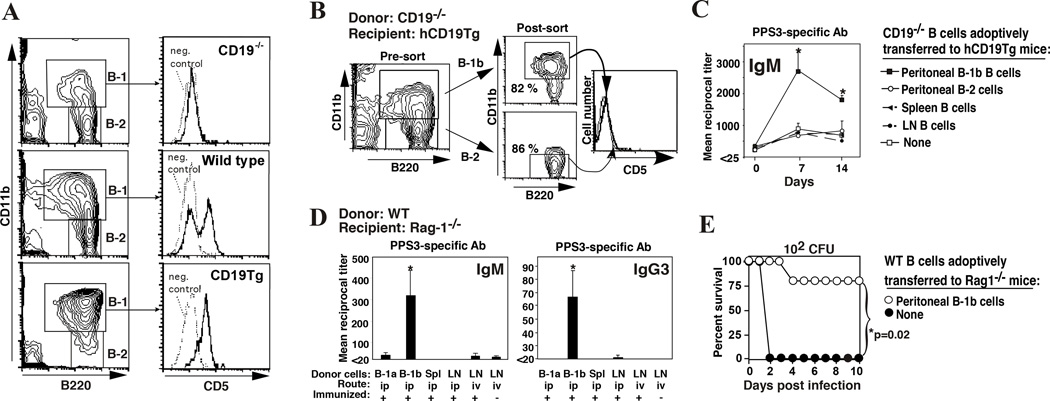
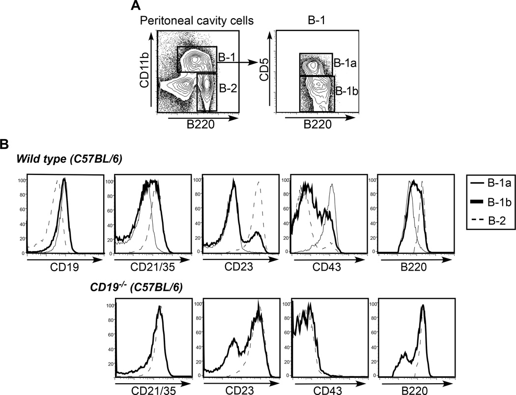
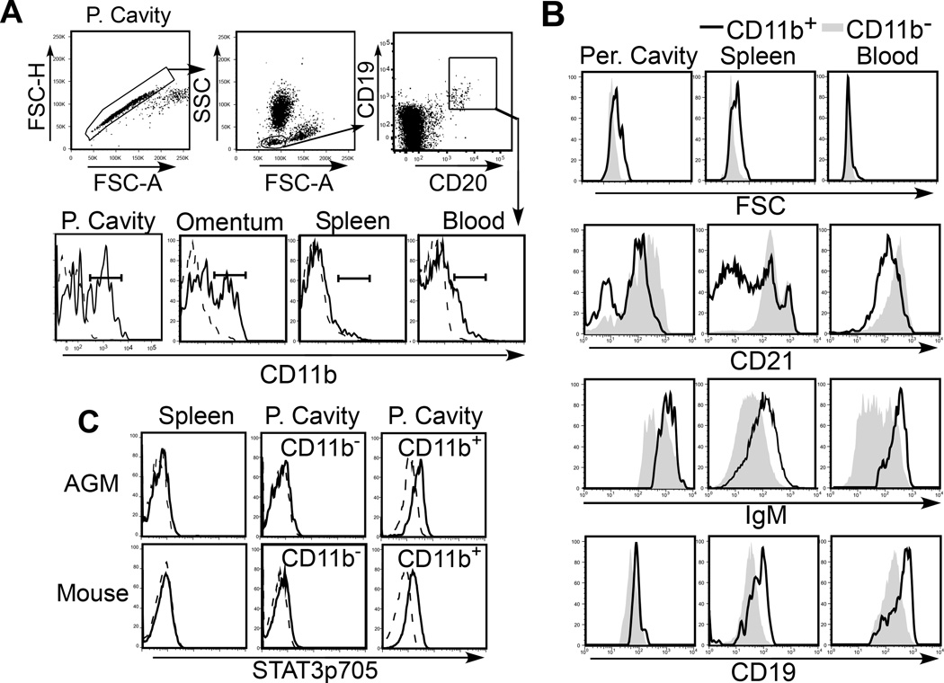
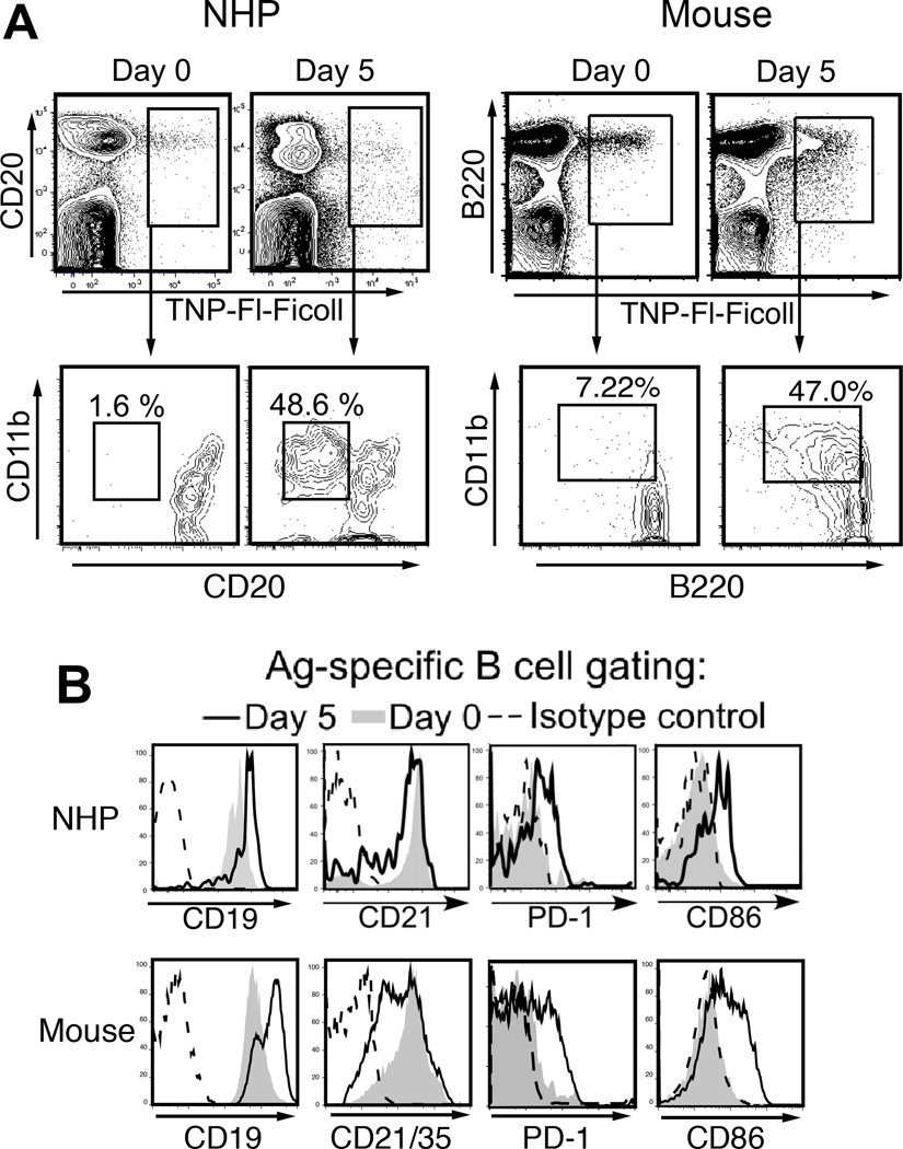
B-1 cells comprise subpopulations of B lymphocytes in mice that display developmental, phenotypic, and functional characteristics that are distinct from those of conventional B cell populations (B-2 cells). Despite the known importance of murine B-1a (CD5(+) ) and B-1b (CD5(-) ) cells in the production of natural antibodies and rapid antigen-specific humoral responses to infection, evidence for B-1 cells in primates, including humans, is very limited. Identifying these cells in humans proves challenging given the limited number of cells that can be obtained from sites expected to harbor increased frequencies of these cells (i.e., peritoneal and pleural cavities) and the need to perform functional analyses on these cells, which, in the case of B-1b cells, must be carried out in vivo. My laboratory has used cynomolgus macaques and African green monkeys to bypass these limitations and to identify and extensively analyze primate B cell populations with the phenotypic and functional characteristics of mouse B-1a and B-1b cells. Our results reveal striking similarities between primate and murine B-1 cells, including a conserved functional role for primate B-1b-like cells in immunity to T cell-independent type 2 antigens.
Keywords: B-1a cells; B-1b cells; T cell-independent type 2 antigens; nonhuman primates.
© 2015 New York Academy of Sciences.
Figures
Similar articles
-
Primate B-1 cells generate antigen-specific B cell responses to T cell-independent type 2 antigens.J Immunol. 2013 Apr 1;190(7):3100-8. doi: 10.4049/jimmunol.1203058. Epub 2013 Mar 1. J Immunol. 2013. PMID: 23455507 Free PMC article.
-
A Comprehensive Atlas of Immunological Differences Between Humans, Mice, and Non-Human Primates.Front Immunol. 2022 Mar 11;13:867015. doi: 10.3389/fimmu.2022.867015. eCollection 2022. Front Immunol. 2022. PMID: 35359965 Free PMC article.
-
Role of B-1a cells in autoimmunity.Autoimmun Rev. 2006 Jul;5(6):403-8. doi: 10.1016/j.autrev.2005.10.007. Epub 2005 Dec 9. Autoimmun Rev. 2006. PMID: 16890894 Review.
-
Characteristics and development of the murine B-1b (Ly-1 B sister) cell population.Ann N Y Acad Sci. 1992 May 4;651:33-43. doi: 10.1111/j.1749-6632.1992.tb24591.x. Ann N Y Acad Sci. 1992. PMID: 1376053 Review.
-
Purification and Immune Phenotyping of B-1 Cells from Body Cavities of Mice.Methods Mol Biol. 2021;2270:27-45. doi: 10.1007/978-1-0716-1237-8_2. Methods Mol Biol. 2021. PMID: 33479891
Cited by
-
Single-cell transcriptome profiling and the use of AID deficient mice reveal that B cell activation combined with antibody class switch recombination and somatic hypermutation do not benefit the control of experimental trypanosomosis.PLoS Pathog. 2021 Nov 11;17(11):e1010026. doi: 10.1371/journal.ppat.1010026. eCollection 2021 Nov. PLoS Pathog. 2021. PMID: 34762705 Free PMC article.
-
Ruxolitinib and oHSV combination therapy increases CD4 T cell activity and germinal center B cell populations in murine sarcoma.Mol Ther Oncol. 2024 Dec 21;33(1):200929. doi: 10.1016/j.omton.2024.200929. eCollection 2025 Mar 20. Mol Ther Oncol. 2024. PMID: 39895689 Free PMC article.
-
B cells in the pneumococcus-infected lung are heterogeneous and require CD4+ T cell help including CD40L to become resident memory B cells.Front Immunol. 2024 Apr 23;15:1382638. doi: 10.3389/fimmu.2024.1382638. eCollection 2024. Front Immunol. 2024. PMID: 38715601 Free PMC article.
-
Chronic Lymphocytic Leukemia B-Cell Normal Cellular Counterpart: Clues From a Functional Perspective.Front Immunol. 2018 Apr 4;9:683. doi: 10.3389/fimmu.2018.00683. eCollection 2018. Front Immunol. 2018. PMID: 29670635 Free PMC article. Review.
-
The widening spectrum of immunological memory.Curr Opin Immunol. 2018 Oct;54:42-49. doi: 10.1016/j.coi.2018.05.013. Epub 2018 Jun 11. Curr Opin Immunol. 2018. PMID: 29902670 Free PMC article. Review.
References
Publication types
MeSH terms
Grants and funding
LinkOut - more resources
Full Text Sources
Other Literature Sources
