. 2015 Jun 12;290(24):14945-62.
doi: 10.1074/jbc.M115.656116. Epub 2015 Apr 30.

p62/Sequestosome-1, Autophagy-related Gene 8, and Autophagy in Drosophila Are Regulated by Nuclear Factor Erythroid 2-related Factor 2 (NRF2), Independent of Transcription Factor TFEB

Affiliations

p62/Sequestosome-1, Autophagy-related Gene 8, and Autophagy in Drosophila Are Regulated by Nuclear Factor Erythroid 2-related Factor 2 (NRF2), Independent of Transcription Factor TFEB

Ashish Jain et al. J Biol Chem. .

Abstract

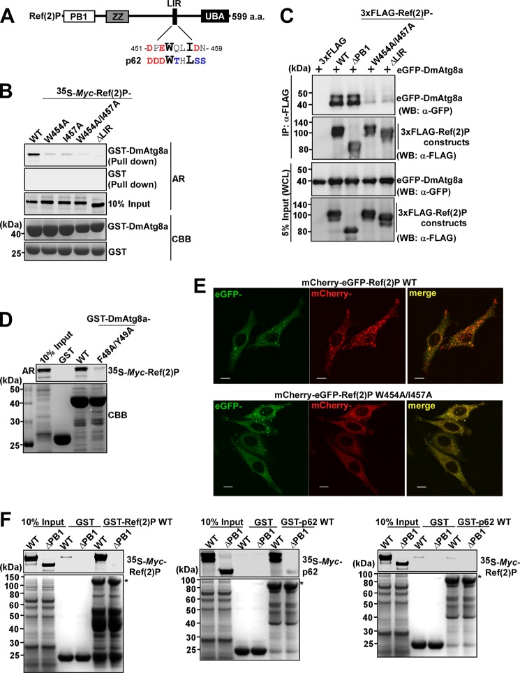
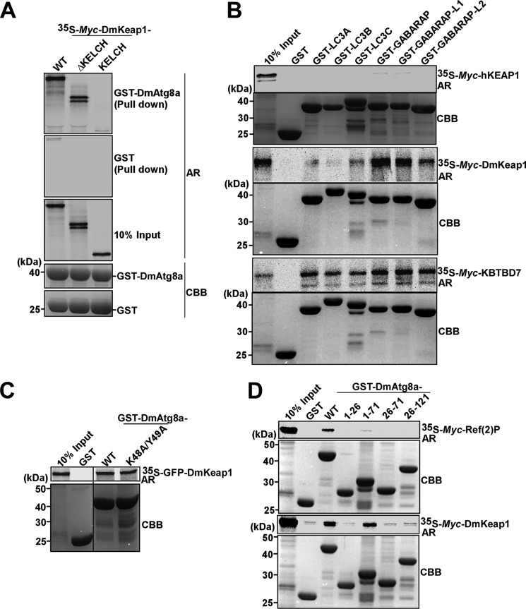
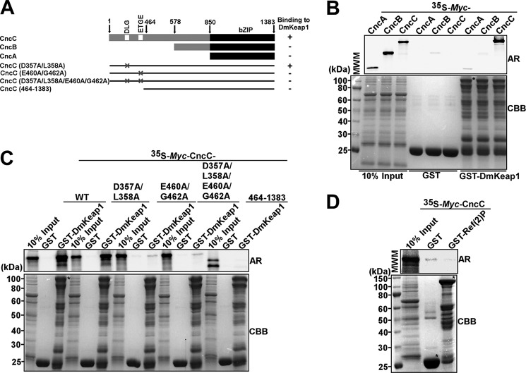
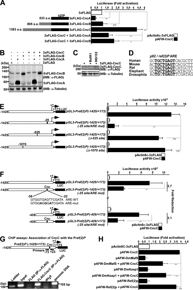
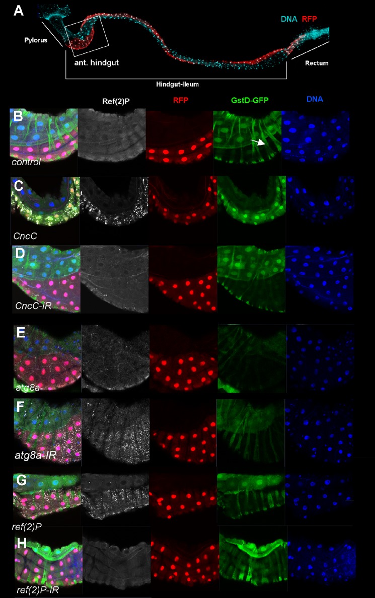
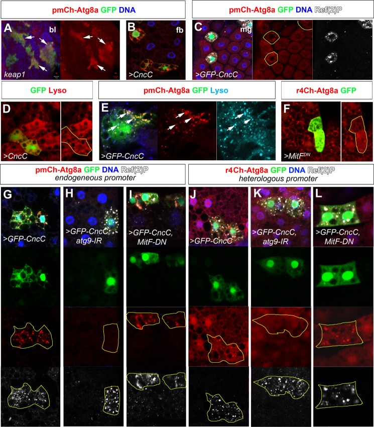
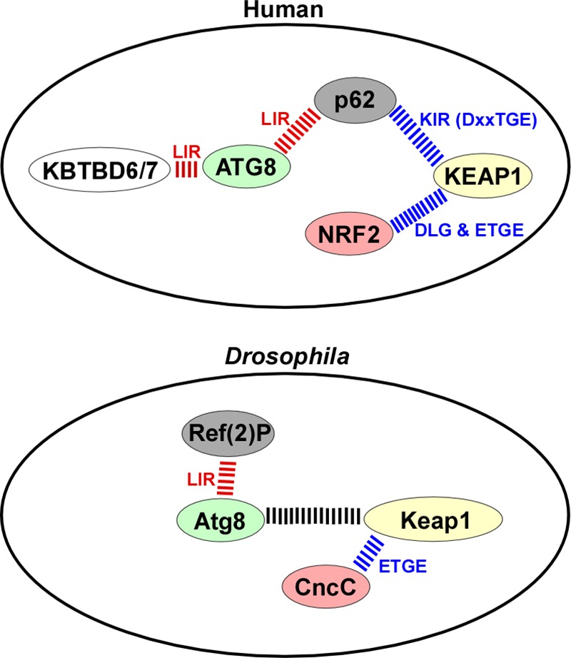
The selective autophagy receptor p62/sequestosome 1 (SQSTM1) interacts directly with LC3 and is involved in oxidative stress signaling in two ways in mammals. First, p62 is transcriptionally induced upon oxidative stress by the NF-E2-related factor 2 (NRF2) by direct binding to an antioxidant response element in the p62 promoter. Second, p62 accumulation, occurring when autophagy is impaired, leads to increased p62 binding to the NRF2 inhibitor KEAP1, resulting in reduced proteasomal turnover of NRF2. This gives chronic oxidative stress signaling through a feed forward loop. Here, we show that the Drosophila p62/SQSTM1 orthologue, Ref(2)P, interacts directly with DmAtg8a via an LC3-interacting region motif, supporting a role for Ref(2)P in selective autophagy. The ref(2)P promoter also contains a functional antioxidant response element that is directly bound by the NRF2 orthologue, CncC, which can induce ref(2)P expression along with the oxidative stress-associated gene gstD1. However, distinct from the situation in mammals, Ref(2)P does not interact directly with DmKeap1 via a KEAP1-interacting region motif; nor does ectopically expressed Ref(2)P or autophagy deficiency activate the oxidative stress response. Instead, DmAtg8a interacts directly with DmKeap1, and DmKeap1 is removed upon programmed autophagy in Drosophila gut cells. Strikingly, CncC induced increased Atg8a levels and autophagy independent of TFEB/MitF in fat body and larval gut tissues. Thus, these results extend the intimate relationship between oxidative stress-sensing NRF2/CncC transcription factors and autophagy and suggest that NRF2/CncC may regulate autophagic activity in other organisms too.

Keywords: autophagy; gene transcription; nuclear factor 2 (erythroid-derived 2-like factor) (NFE2L2) (Nrf2); oxidative stress; p62 (sequestosome 1 (SQSTM1)).


Figures

FIGURE 1.
FIGURE 1.
Drosophila p62, Ref(2)P, interacts with DmAtg8a. A, schematic map of Ref(2)P with PB1, ZZ, and UBA domains, including a putative LIR-motif. The human p62 LIR motif is shown below the Ref(2)P LIR motif for comparison. B, Ref(2)P WT binds DmAtg8a, whereas indicated point mutations or deletion of the LIR motif in Ref(2)P completely abolishes the DmAtg8a binding. Myc-tagged constructs in vitro translated in the presence of [35S]methionine were analyzed for binding in GST pull-down assays. Bound proteins were detected by autoradiography (AR). Bottom panels, a Coomassie-stained gel of immobilized GST or GST-tagged proteins (CBB). C, eGFP-DmAtg8a was co-immunoprecipitated (IP) with FLAG-Ref(2)P WT and ΔPB1 but not with the indicated point mutated or deleted constructs of the LIR-motif from S2R+ cell extracts. Cells were co-transfected with the indicated FLAG-Ref(2)P constructs and eGFP-DmAtg8a and immunoprecipitated with anti-FLAG antibody 72 h after transfection. Precipitated proteins were detected by Western blotting (WB) using the indicated antibodies. WCL, whole cell lysate. D, DmAtg8a WT binds to Ref(2)P, whereas the F48A/Y49A LIR-binding site mutation abolishes Ref(2)P binding. E, the LIR motif of Ref(2)P is required for its accumulation in acidic vesicles. HeLa cells were transfected with mCherry-eGFP-Ref(2)P (WT or W454A/I457A), and the accumulation in acidic vesicles (red only) was analyzed 18 h after transfection. Scale bars, 10 μm. F, Ref(2)P binds to itself via its PB1 domain, but it does not interact with p62. Full-length proteins are indicated with asterisks.
FIGURE 2.
FIGURE 2.
Ref(2)P does not interact directly with DmKeap1 via a KIR motif. A, Ref(2)P does not interact with DmKeap1. Myc-tagged constructs were in vitro translated and analyzed for binding in pull-down assays. Full-length proteins are indicated with asterisks. B, sequence alignment of p62 orthologs from representative metazoan species showing the LIR and KIR motifs boxed. C, schematic map of Ref(2)P indicating different deletion constructs employed in D, E, and F to map the ubiquitin-mediated association with DmKeap1 in vivo. D, eGFP-Ref(2)P WT, -ΔPB1, and W454A/I457A (LIR mutant) but not the ΔUBA construct were co-immunoprecipitated with FLAG-DmKeap1 from S2R+ cell extracts. Cells were co-transfected with the indicated constructs. E and F, eGFP-DmKeap1 and ubiquitinated proteins were co-immunoprecipitated with FLAG-Ref(2)P WT from S2R+ cell extracts but not with the ΔUBA construct or a point mutant in the UBA domain (M565V). G, four ubiquitinated lysines were detected by MS/MS analysis of eGFP-DmKeap1 immunoprecipitated from S2R+ cell extracts with anti-GFP antibody 72 h after transfection. CBB, Coomassie Brilliant Blue; AR, autoradiography; WB, Western blot; IP, immunoprecipitation; WCL, whole cell lysate.
FIGURE 3.
FIGURE 3.
DmKeap1 interacts with DmAtg8a. A, full-length DmKeap1 and ΔKELCH constructs of DmKeap1 interact with DmAtg8a, whereas KELCH alone does not. B, DmKeap1 and KBTBD7 interact with the human ATG8 proteins (middle and bottom), whereas hKEAP1 does not (top). Myc-tagged constructs were in vitro translated and tested for binding. C, both the WT and the F48A/Y49A LIR-binding site mutant of DmAtg8a bind equally well to DmKeap1. D, DmAtg8a(1–71) interacts with DmKeap1 but not with Ref(2)P. CBB, Coomassie Brilliant Blue; AR, autoradiography.
FIGURE 4.
FIGURE 4.
DmKeap1 levels are affected by programmed but not basal autophagy. A, L3 larval extracts of the following genotypes were immunoblotted against DmKeap1: w1118 (control), atg61, atg8d4, atg13D81, ref(2)P03993, tubulin-Gal4/+;UAS-GFP-DmKeap1/+, or tubulin-Gal4/+;DmKeap1/+. The full-length DmKeap1 is indicated with an asterisk. B, large atg13 mutant clones of the wing imaginal disc strongly accumulated Ref(2)P, indicating that basal autophagy was compromised, whereas ubiquitously expressed GFP-DmKeap1 remained uniform between adjacent mutant/control cell borders (atg13 mutant clones are indicated by yellow outline, RFP-negative). C, schematic depicting midgut development with EC growing substantially in size by endoreplication during the L3 larval stage and adult midgut progenitor (AMP) cell clusters increasing from 1 to 9–12 cells by the wL3 stage. In the white prepupal stage (WPP), EC cells shrink by autophagy before being replaced by AMPs. D and F, DmKeap1 and GFP-DmKeap1 localized to cytoplasmic structures in EC and AMP cells in mid L3 midguts. E and G, by the wL3 stage, DmKeap1 and GFP-DmKeap1 are reduced in EC relative to AMP cells. H, co-expression of GFP-DmKeap1 and Ch-Ref(2)P in midgut cells of L3 larvae showed co-localization but not general overlap between GFP-DmKeap1 and Ch-Ref(2)P structures (Pearson coefficient 0.57). I, starvation of mL3 larvae led to a robust induction of Ch-DmAtg8a structures with partial overlap with Ref(2)P (PC = 0.28), indicating autolysosomes. No extensive overlap was observed between GFP-DmKeap1 and Ref(2)P (PC = 0.05) or Ch-Atg8a (PC = 0.15). J, atg13 mutant EC cells, identifiable by the lack of RFP, remained large in the white prepupal stage (yellow outline). GFP-DmKeap1 intensities were increased in atg13 mutant EC cells versus neighboring autophagy proficient cells undergoing cytoplasmic shrinkage. K, in late wandering L3 stage larvae, autophagy-proficient control cells were significantly smaller (mean relative cell size 0.57 ± 0.07 (S.E.)) and atg13 mutant cells displayed a clear and consistent increase in GFP-Keap1 levels relative to control cells mutant cells (3.8 ± 0.4-fold (S.E.) increase in GFP-Keap1 levels). L, relative pairwise comparison of atg13 mutant and neighboring control cells for GFP-Keap1 levels and cell size (n = 27 cells, pairwise comparisons from three animals). Genotypes were w1118 (D and E), y,w,hs-flp/w*;UAS-GFP-DmKeap1;act-Gal4;FRT82,atg13d81/FRT82,RFP (B and J), UAS-GFP-DmKeap1/+;tubulin-Gal4 (F and G), UAS-GFP-DmKeap1/+;tubulin-Gal4,UAS-Cherry-Ref(2)P/+ (H), and UAS-GFP-DmKeap1/+;tubulin-gal4/pmCherry-Atg8a (I). eL3, early L3 stage; mL3, mid-L3 stage; wL3, wandering L3 stage. WB, Western blot. Error bars, S.E.
FIGURE 5.
FIGURE 5.
DmKeap1 interacts with CncC. A, schematic map of Cnc indicating the different isoforms employed in B and mutant constructs used in C to map the interaction with DmKeap1. B, only the CncC isoform of Cnc interacts with DmKeap1. C, CncC interacts with DmKeap1 via its ETGE motif. In B and C, the depicted Myc-tagged constructs were in vitro translated and used in pull-down assays. D, Ref(2)P does not interact with CncC in vitro. The full-length proteins are indicated with an asterisk. CBB, Coomassie Brilliant Blue; AR, autoradiography.
FIGURE 6.
FIGURE 6.
Mapping of a CncC binding site in the ref(2)P promoter. A, CncC, but not CncA or CncB, transactivates the ref(2)P promoter. S2R+ cells were co-transfected with an empty vector (pActin5C-3xFLAG) or the indicated Cnc constructs (75 ng each) together with pGL3-Pref(2)P(-1425/+173) (150 ng). Cells were harvested 48 h after transfection. B, ectopically expressed CncC is not detectable by Western blot under normal conditions. S2R+ cells were transfected with the indicated Cnc constructs and extracts analyzed by Western blot using anti-FLAG antibody 48 h after transfection. α-Tubulin was used as a loading control. C, ectopically expressed CncC is degraded by the proteasome because it was stabilized by treatment for 6 h before harvesting with 25 μm MG132 but not 0.2 μm BafA1. α-Tubulin was used as loading control. D, the ARE in ref(2)P promoter is conserved in human, mouse, rat, elephant, and D. melanogaster. The conserved residues in the core ARE consensus are shown in boldface lettering. E, the ARE at position −25 mediates CncC-derived induction of the ref(2)P promoter. Reporter gene assays were performed on the indicated (WT, mutated, or deleted) ref(2)P(−1425/+173) promoter constructs. S2R+ cells were co-transfected with the indicated constructs and harvested 72 h after transfection. F, CncC activates the ref(2)P promoter via the ARE motif at the −25 position. S2R+ cells were co-transfected with the indicated constructs and harvested 48 h after transfection. For each ref(2)P promoter construct, -fold reduction caused by mutation in the ARE element is shown to the right. G, CncC is associated with the ref(2)P promoter in vivo. S2R+ cells were co-transfected with either pGL3-Pref(2)P(−1425/+173) and pActin5C-3xFLAG or pGL3-Pref(2)P(−1425/+173) and pAFW-CncC (5 μg of each plasmid per 8 × 10⁶ S2R+ cells), and cell extracts were prepared 72 h after transfection. Immunoprecipitation was done with anti-FLAG antibody. PCR analysis of the immunoprecipitated chromatin was carried out using primers flanking the ARE (positions −73 and +23, respectively). Genomic DNA was used as a positive PCR control, and distilled H2O was used as a no template control. H, Ref(2)P does not induce its own promoter, whereas DmKeap1 and DmMafS repress CncC-mediated ref(2)P promoter activity. S2R+ cells were co-transfected with the indicated constructs and harvested 48 h after transfection. The data shown are the mean ± S.D. activities obtained in one experiment performed in triplicate and are representative of four independent experiments (ns, not significant; *, p < 0.05; **, p < 0.01 compared with the controls as shown). Error bars, S.E. WB, Western blot.
FIGURE 7.
FIGURE 7.
CncC induces Ref(2)P expression and an oxidative stress response associated gene in D. melanogaster. A, anatomical overview of the larval hindgut (ileum) with the anterior pylorus and posterior rectum. The dorsal expression domain of engrailed driving RFP is shown together with a nuclear stain (DNA). B–H, single confocal sections of the anterior hindgut (boxed in A) with nuclei, Ref(2)P protein, transgene expression domain (RFP), and the gstD-GFP oxidative stress reporter. B, control animal showing the normal slightly higher expression level of GstD-GFP in the non-en-Gal4, UAS-RFP-expressing cells. This difference is seen also when engrailed-UAS-RFP is not present (data not shown). Transverse GFP-positive stripes are cytoplasmic signal from circular muscles outside of the gut endothelium (arrow). Expression of CncC (C), but not CncC knockdown by RNAi (CncC-IR) (D), led to a strong induction of Ref(2)P protein levels and GstD-GFP expression. Neither overexpression of DmAtg8a (E) nor DmAtg8a knockdown (F) affected oxidative stress levels, but a strong accumulation of Ref(2)P protein was observed, indicating that autophagy was effectively blocked. Ref(2)P overexpression (G) or ref(2)P knockdown (H) had no effect on oxidative stress levels, as judged by GstD-GFP-expression. Genotypes were as follows: gstD-GFP;en-Gal4,UAS-RFP/CyO (A and B), gstD-GFP/+;en-Gal4,UAS-RFP/+;UAS-CncC (C), gstD-GFP/+;en-Gal4,UAS-RFP/+;CncC-IRTRIP.HMS00650/+ (D), gstD-GFP/UAS-atg8aEP362;en-Gal4,UAS-RFP/+ (E), gstD-GFP/+;en-Gal4,UAS-RFP/+;atg8a-IRTRIP.JF02895/+ (F), gstD-GFP/+;en-Gal4,UAS-RFP/+;UAS-ref(2)P-5/+ (G), and gstD-GFP/+;en-Gal4,UAS-RFP/+;UAS-Ref(2)P-IR/+ (H).
FIGURE 8.
FIGURE 8.
CncC induces autophagy independent of MitF. A, large GFP-labeled keap1 loss of function clones in the larval brain lobe accumulate mCherry-Atg8a expressed under its endogenous upstream regulatory sequence (pmCh-Atg8a). B–E, clonal overexpression of CncC and GFP-CncC in Drosophila fat body (fb) (B) and midgut (mg) (C) cells similarly induces pmCherry-Atg8a up-regulation and formation of cytoplasmic puncta partially co-localizing with Ref(2)P. D and E, CncC or GFP-CncC overexpression in fat body clones induces Lysotracker-positive structures partially co-localizing with mCherry-Atg8a. F, clonal expression of a dominant negative version of the Drosophila TFEB orthologue, MitF, inhibits mCherry-Atg8a accumulation upon oxidative stress-induced autophagy. G–I, clonal GFP-CncC expression in fat body cells leads to up-regulation and accumulation or mCherry-Atg8a and Ref(2)P. Punctate accumulation of mCherry-Atg8a, but not Ref(2)P, is reversed upon RNAi-mediated depletion of atg9a, whereas both are unaffected by expression of dominant negative MitF. J–L, another mCherry-Atg8a reporter expressed under a synthetic fat body promoter, r4ChAtg8a, similarly shows partial co localization of mCherry-Atg8a and Ref(2)P but no up-regulation of mCherry-Atg8a levels upon GFP-CncC expression. Autophagy induction but not Ref(2)P accumulation is dependent on atg9, whereas reducing MitF function has no effect. Genotypes: (A) y,w, ey-flp/+; act>CD2>GAL4, UAS-GFP/pmCh-Atg8a;FRT82, keap1D036/FRT82,tub-Gal80 (A); hsp70-flp/+ (B); pmChAtg8a/+; act>CD2>GAL4, UAS-GFPnls/UAS-CncC (C, E, and G); hsp70-flp/+; UAS-CncC/+; act>CD2>GAL4, UAS-GFPnls/+ (D); hsp70-Flp/+; UAS-Dicer/+; r4-mCh::Atg8a, act>CD2>GAL4, UAS-GFPnls/UAS-MitF-DN (F); hsp70-flp/+; pmChAtg8a/UAS-GFP-CncC; act>CD2>GAL4, UAS-GFPnls/UAS-atg9-IR (H); hsp70-flp/+; pmChAtg8a/UAS-GFP-CncC; act>CD2>GAL4, UAS-GFPnls/UAS-MitF-DN (I); hsp70-Flp/+; UASDicer/UAS-GFP-CncC; r4-mCh::Atg8a, act>CD2>GAL4, UAS-GFPnls/+ (J); hsp70-Flp/+; UASDicer/UAS-GFP-CncC; r4-mCh::Atg8a, act>CD2>GAL4, UAS-GFPnls/UAS-atg9-IR (K); hsp70-Flp/+; UAS-Dicer/UAS-GFP-CncC; r4-mCh::Atg8a, act>CD2>GAL4, UAS-GFPnls/UAS-MitF-DN (L).
FIGURE 9.
FIGURE 9.
Comparative protein-protein interaction summary for the human and D. melanogaster proteins studied. The LIR-mediated interactions are indicated in red, and those mediated by DLG and ETGE-like motifs are shown in blue. The exact nature and role of the interaction between Atg8a and DmKeap1 is not known.

Similar articles

Cited by

References

    1. Mathers J., Fraser J. A., McMahon M., Saunders R. D., Hayes J. D., McLellan L. I. (2004) Antioxidant and cytoprotective responses to redox stress. Biochem. Soc. Symp. 157–176 - PubMed
    1. Hayes J. D., McMahon M. (2009) NRF2 and KEAP1 mutations: permanent activation of an adaptive response in cancer. Trends Biochem. Sci. 34, 176–188 - PubMed
    1. Nguyen T., Nioi P., Pickett C. B. (2009) The Nrf2-antioxidant response element signaling pathway and its activation by oxidative stress. J. Biol. Chem. 284, 13291–13295 - PMC - PubMed
    1. Taguchi K., Motohashi H., Yamamoto M. (2011) Molecular mechanisms of the Keap1-Nrf2 pathway in stress response and cancer evolution. Genes Cells 16, 123–140 - PubMed
    1. McMahon M., Itoh K., Yamamoto M., Chanas S. A., Henderson C. J., McLellan L. I., Wolf C. R., Cavin C., Hayes J. D. (2001) The Cap'n'Collar basic leucine zipper transcription factor Nrf2 (NF-E2 p45-related factor 2) controls both constitutive and inducible expression of intestinal detoxification and glutathione biosynthetic enzymes. Cancer Res. 61, 3299–3307 - PubMed

Publication types

LinkOut - more resources