. 2015 Jun 19;290(25):15697-15706.
doi: 10.1074/jbc.M115.650176. Epub 2015 Apr 30.

The Helicase Activity of Ribonuclease R Is Essential for Efficient Nuclease Activity

Affiliations

The Helicase Activity of Ribonuclease R Is Essential for Efficient Nuclease Activity

Sk Tofajjen Hossain et al. J Biol Chem. .

Abstract

RNase R, which belongs to the RNB family of enzymes, is a 3' to 5' hydrolytic exoribonuclease able to digest highly structured RNA. It was previously reported that RNase R possesses an intrinsic helicase activity that is independent of its ribonuclease activity. However, the properties of this helicase activity and its relationship to the ribonuclease activity were not clear. Here, we show that helicase activity is dependent on ATP and have identified ATP-binding Walker A and Walker B motifs that are present in Escherichia coli RNase R and in 88% of mesophilic bacterial genera analyzed, but absent from thermophilic bacteria. We also show by mutational analysis that both of these motifs are required for helicase activity. Interestingly, the Walker A motif is located in the C-terminal region of RNase R, whereas the Walker B motif is in its N-terminal region implying that the two parts of the protein must come together to generate a functional ATP-binding site. Direct measurement of ATP binding confirmed that ATP binds only when double-stranded RNA is present. Detailed analysis of the helicase activity revealed that ATP hydrolysis is not required because both adenosine 5'-O-(thiotriphosphate) and adenosine 5'-(β,γ-imino)triphosphate can stimulate helicase activity, as can other nucleoside triphosphates. Although the nuclease activity of RNase R is not needed for its helicase activity, the helicase activity is important for effective nuclease activity against a dsRNA substrate, particularly at lower temperatures and with more stable duplexes. Moreover, competition experiments and mutational analysis revealed that the helicase activity utilizes the same catalytic channel as the nuclease activity. These findings indicate that the helicase activity plays an essential role in the catalytic efficiency of RNase R.

Keywords: ATP; Escherichia coli (E. coli); RNA degradation; RNA helicase; ribonuclease.


Figures

FIGURE 1.
FIGURE 1.
A, schematic showing the domain organization of E. coli RNase R. The three RNA-binding domains CSD1, CSD2, and S1, the catalytic nuclease domain, and the C-terminal basic domain are shown. The position of the Walker A and Walker B motifs are noted. B, sequence alignment using UniProt of available RNase R sequences from 49 different genera of mesophilic bacteria that show conservation of the ATP-binding Walker A and Walker B motifs, highlighted in yellow.
FIGURE 2.
FIGURE 2.
Helicase activity of RNase R. A, effect of ATP on helicase activity of RNase R and RNase R mutant proteins. Assays were carried out as described under “Experimental Procedures” with 2.5 nm [5′-32P]U12C5:G5A29 substrate (shown on left) and 1 μm enzyme. B, effect of ATP concentration. Activity was measured using 1 μm RNase R D272N as in panel A at the indicated ATP concentrations for 30 min. C is a no enzyme control incubated for 30 min. Shown is a representative experiment carried out three times with essentially identical results.
FIGURE 3.
FIGURE 3.
Helicase activity of RNase R D272N in the presence of ATP or its nonhydrolyzable ATP analogues. Assays were carried out as described under “Experimental Procedures” using 2 mm ATP or ATP analogs, 2.5 nm [5′-32P]U12C5:G5A29 (shown on right) and 0.1 μm RNase R D272N. Samples were incubated for 30 min. C is a no enzyme control incubated for 30 min. Shown is a representative experiment. The quantification presented below each gel is the average of four experiments.
FIGURE 4.
FIGURE 4.
Effect of helicase activity on RNase R nuclease activity against single-stranded RNA. A, exoribonuclease activity of wild type RNase R and helicase mutant proteins on a single-stranded substrate. Assays were carried out as described under “Experimental Procedures” for the indicated times at 37 °C in the presence of 5 mm ATP. The number below each lane is the percent substrate remaining. Shown is a representative experiment carried out three times. B, effect of ATP on hydrolysis of single-stranded RNA at different temperatures. Assays were carried out in reaction mixtures containing 50 mm Tris-HCl (pH 8.0), 300 mm KCl, 0.25 mm MgCl2, 5 mm DTT, 10 μm ss17-A17 oligoribonucleotide substrate, and 0.025 μm of enzyme, with and without 5 mm ATP. Portions were taken at the indicated times and analyzed by denaturing PAGE followed by autoradiography. The quantification presented below is the average of three experiments ± S.D. Δ, −ATP; □, +ATP.
FIGURE 5.
FIGURE 5.
Effect of helicase activity on RNase R nuclease activity against double-stranded RNA. A, exoribonuclease activity of wild type RNase R on ds17-A17 (G5A29:U12C5) in the presence or absence of 5 mm ATP at different temperatures. B, exoribonuclease activity of RNase R K736A mutant protein on a substrate containing ds17-A17 (G5A29:U12C5) in the presence or absence of 5 mm ATP at different temperatures. C, exoribonuclease activity of wild type RNase R on GC-rich ds17-A17 (G10A24:U7C10) in the presence or absence of 5 mm ATP at different temperatures. All assays were carried out in reaction mixtures containing 50 mm Tris-HCl (pH 8.0), 300 mm KCl, 0.25 mm MgCl2, 5 mm DTT, 10 μm oligoribonucleotide substrate, and 0.25 μm enzyme. Portions were taken at the indicated times and analyzed by denaturing PAGE followed by autoradiography. The quantification shown is the average of three experiments ± S.D. Δ, −ATP; □, +ATP.
FIGURE 6.
FIGURE 6.
Duplex RNA stimulates ATP binding. ATP binding in the presence of 2 mm ATP was determined using a filter binding assay as described under “Experimental Procedures.” Varying amounts of single-stranded (G5A29) or double-stranded (G5A29:U12C5) RNA were added as indicated in the figure. The amount of bound ATP in the absence of both enzyme and RNA was subtracted from each value. The quantification shown is the average of two experiments ± S.D. Data are presented as the percent of RNase R molecules with bound ATP assuming a single ATP molecule binds per molecule of RNase R.
FIGURE 7.
FIGURE 7.
Helicase activity utilizes the nuclease catalytic channel. A, helicase activity of RNase R D272N in the presence of single-stranded oligonucleotides. Assays were carried out for 30 min in the presence of different concentrations of A4 or A17, as indicated. B, helicase activity of RNase R catalytic channel mutants. Helicase assays in both panels were carried out in a reaction mixture containing 2.5 nm RNA substrate, 0.1 μm enzyme, and 2 mm ATP for the indicated times. Left panel, ○, RNase R D272N; □, RNase R D272N,R572K; right panel, ○, RNase R D272N; □, RNase R D272N,H456N,H565T,R572K. Quantification shown is the average of three experiments ± S.D. A representative gel is shown for each experiment.
FIGURE 8.
FIGURE 8.
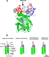
Structural and schematic model of RNase R based on the closely related RNase II. A, approximate locations of the Walker A and Walker B motifs (highlighted by ovals) shown on the ribbon structure of the closely related RNase II crystallized with an RNA fragment trapped in the nuclease channel (21). The view shown is from the front of the enzyme, with the RNA binding clamp on the top (N-terminal CSD1 and CSD2 domains in pink and blue, and the C-terminal S1 domain in red). These three domains form an open-ended RNA-binding funnel that leads into the narrow channel in the nuclease domain (shown in green). The RNA strand is shown in a stick representation, and one of the Asp residues in Walker B (D102 in RNase II) can be seen as blue sticks at the bottom of the oval. B, schematic model of single-strand and double-strand RNA bound on RNase R to explain enhancement of nuclease activity in the presence of ATP. The RNA-binding domains are shown in pink, blue, and red for the CSD1, CSD2, and S1 domains, respectively. The nuclease domain is in green with its catalytic center highlighted by a pink arrow. No change in structure occurs upon single-strand RNA binding. Upon double-strand RNA binding, the Walker A and Walker B motifs come close to each other to generate an ATP-binding site. ATP binding stimulates strand separation, likely due to a conformational change in the protein. The newly separated strand moves forward into the nuclease channel to position the next nucleotide for cleavage at the 3′ end. The color coding in panels A and B is the same.

Similar articles

Cited by

References

    1. Cheng Z. F., Deutscher M. P. (2005) An important role for RNase R in mRNA decay. Mol. Cell 17, 313–318 - PubMed
    1. Vincent H. A., Deutscher M. P. (2006) Substrate recognition and catalysis by the exoribonuclease RNase R. J. Biol. Chem. 281, 29769–29775 - PubMed
    1. Vincent H. A., Deutscher M. P. (2009) The roles of individual domains of RNase R in substrate binding and exoribonuclease activity: the nuclease domain is sufficient for digestion of structured RNA. J. Biol. Chem. 284, 486–494 - PMC - PubMed
    1. Vincent H. A., Deutscher M. P. (2009) Insights into how RNase R degrades structured RNA: analysis of the nuclease domain. J. Mol. Biol. 387, 570–583 - PMC - PubMed
    1. Cheng Z. F., Deutscher M. P. (2003) Quality control of ribosomal RNA mediated by polynucleotide phosphorylase and RNase R. Proc. Natl. Acad. Sci. U.S.A. 100, 6388–6393 - PMC - PubMed

Publication types

MeSH terms

LinkOut - more resources