. 2015 Jun 26;290(26):16099-115.
doi: 10.1074/jbc.M115.659318. Epub 2015 Apr 30.

MicroRNA-193a-3p Reduces Intestinal Inflammation in Response to Microbiota via Down-regulation of Colonic PepT1

Affiliations

MicroRNA-193a-3p Reduces Intestinal Inflammation in Response to Microbiota via Down-regulation of Colonic PepT1

Xin Dai et al. J Biol Chem. .

Abstract

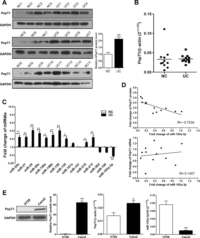
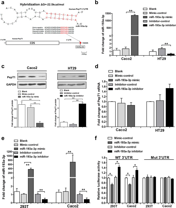
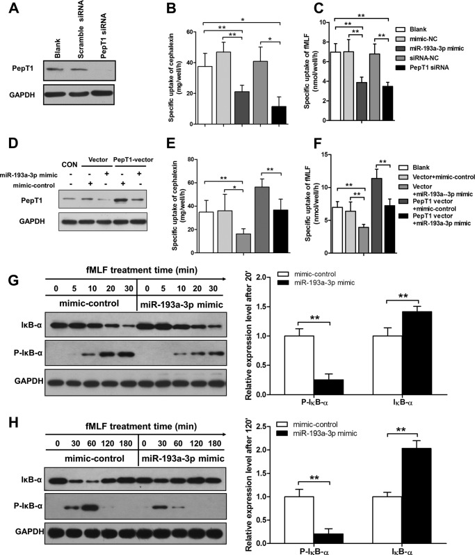
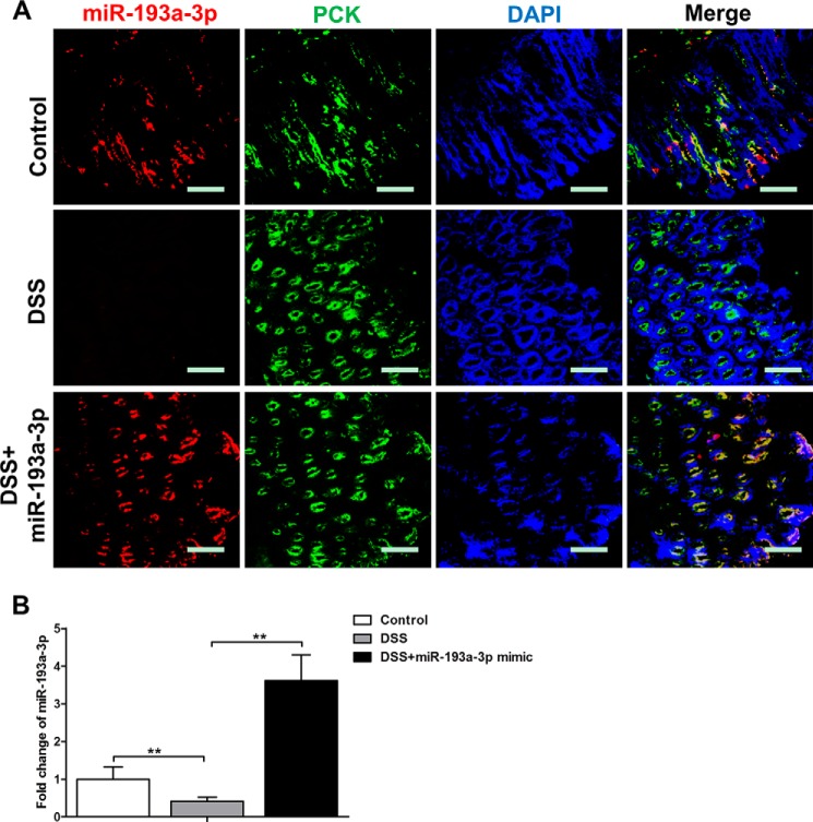
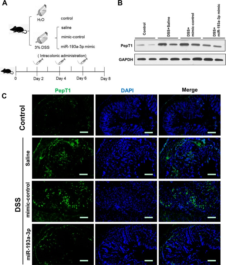
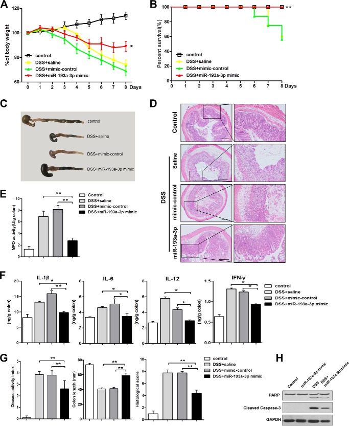
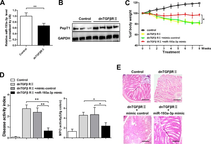
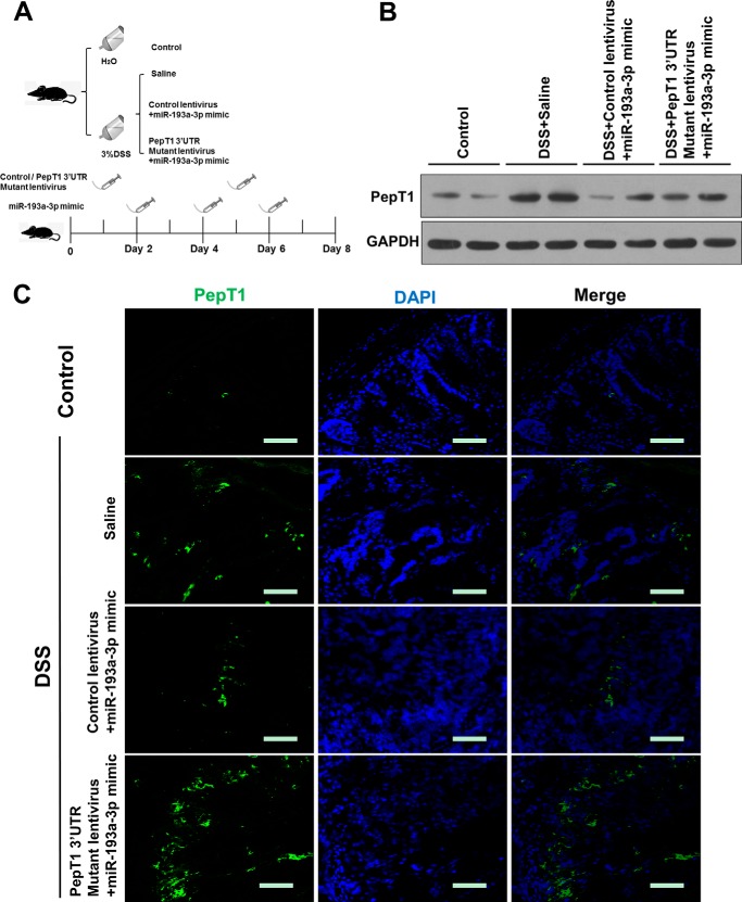
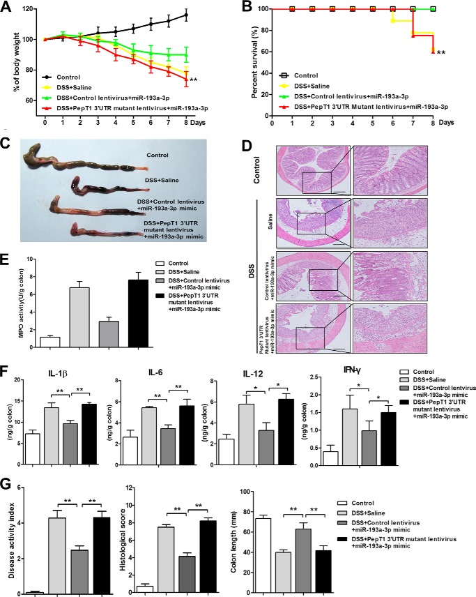
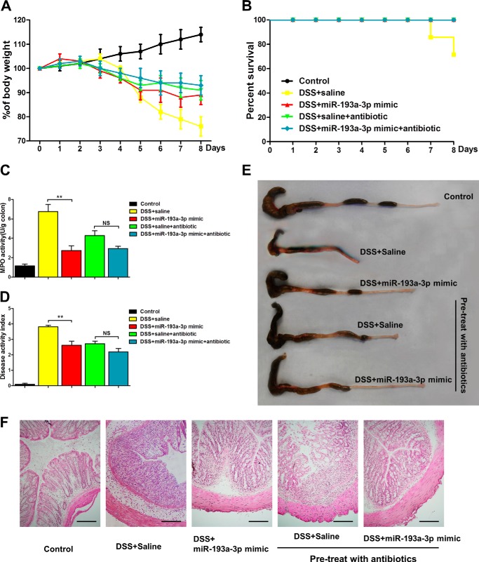
Intestinal inflammation is characterized by epithelial disruption, leading to the loss of barrier function, recruitment of immune cells, and host immune responses to gut microbiota. PepT1, a di/tripeptide transporter that uptakes bacterial products, is up-regulated in inflamed colon tissue, which implies its role in bacterium-associated intestinal inflammation. Although microRNA (miRNA)-mediated gene regulation has been found to be involved in various processes of inflammatory bowel disease (IBD), the biological function of miRNAs in the pathogenesis of IBD remains to be explored. In this study we detected miRNA expression patterns in colon tissues during colitis and investigated the mechanism underlying the regulation of colonic PepT1 by miRNAs. We observed an inverse correlation between PepT1 and miR-193a-3p in inflamed colon tissues with active ulcerative colitis, and we further demonstrated that miR-193a-3p reduced PepT1 expression and activity as a target gene and subsequently suppressed the NF-κB pathway. Intracolonic delivery of miR-193a-3p significantly ameliorated dextran sodium sulfate-induced colitis, whereas the overexpression of colonic PepT1 via PepT1 3'-untranslated region mutant lentivirus vector abolished the anti-inflammatory effect of miR-193a-3p. Furthermore, antibiotic treatment eliminated the difference in the dextran sodium sulfate-induced inflammation between the presence and absence of miR-193a-3p. These findings suggest that miR-193a-3p regulation of PepT1 mediates the uptake of bacterial products and is a potent mechanism during the colonic inflammation process. Overall, we believe miR-193a-3p may be a potent regulator of colonic PepT1 for maintaining intestinal homeostasis.

Keywords: gene regulation; inflammatory bowel disease (IBD); microRNA (miRNA); mucosal immunology; peptide transport.


Figures

FIGURE 1.
FIGURE 1.
The inverse correlation between PepT1 and miR-193a-3p. A, Western blotting of PepT1 levels in active UC and normal control (NC) colon tissues: representative Western blot (left panels) and quantitative analysis of PepT1 levels (right panels). B, The relative expression of PepT1 mRNA transcripts in active UC and NC tissues by qRT-PCR. C, the relative miRNA expression ratios of active UC to NC tissues. D, Pearson's correlation scatter plots of the -fold changes of miR-193a-3p and PepT1 mRNA and PepT1 protein. E, PepT1 protein expression by Western blotting and PepT1 mRNA and miR-193a-3p expression by qRT-PCR in Caco2 and HT29 cells. *, p < 0.05; **, p < 0.01.
FIGURE 2.
FIGURE 2.
Identification of PepT1 as a target of miR-193a-3p. A, schematic description of the conserved binding site for miR-193a-3p and human PepT1. B, qRT-PCR analysis of miR-193a-3p expression levels after transfection with the miR-193a-3p mimic, mimic-control in Caco2, the miR-193a-3p inhibitor, and inhibitor-control in HT29 cells normalized to U6. C, Western blotting of PepT1 protein expression levels after above transfection in Caco2 cells and HT29 cells. D, qRT-PCR analysis of PepT1 mRNA levels after above transfection in Caco2 and HT29 cells. E, qRT-PCR analysis of miR-193a-3p expression levels after transfection with the miR-193a-3p mimic, mimic-control, the miR-193a-3p inhibitor, and inhibitor-control in 293T and Caco2 cells. F, luciferase reporter activity after expression of the above transfections in 293T cells. Luciferase reporters carrying the wild-type (WT) or mutant (Mut) PepT1 3′-UTR were cotransfected into 293T and Caco2 cells along with the indicated oligonucleotides. *, p < 0.05; **, p < 0.01.
FIGURE 3.
FIGURE 3.
MiR-193a-3p reduces PepT1 transport ability and suppresses the bacterial peptide induced inflammatory response in vitro. A, PepT1 siRNA compared with the scrambled siRNA in Caco2 cells. B and C, PepT1 transport activity 24 h after transfection with the miR-193a-3p mimic or PepT1 siRNA expressed by measuring the specific uptake of cephalexin and fMLF. D, Western blotting of PepT1 protein expression levels after cotransfection of PepT1 overexpressing-vector and miR-193a-3p mimic or mimic-control. E and F, PepT1 transport activity 24 h after the cotransfection above expressed by measuring the specific uptake of cephalexin and fMLF. G and H, Caco2 cells pretransfected with the miR-193a-3p mimic or mimic control were stimulated with fMLP (100 nm) for the indicated times, and levels of phosphor IκB-α and IκB-α were measured by Western blotting. Bar graphs represent the densitometric quantification at the 30- and 120-min time points. *, p < 0.05; **, p < 0.01.
FIGURE 4.
FIGURE 4.
Cellular location of miR-193a-3p after miR-193a-3p mimic administration in vivo. A, immunofluorescent staining with CEC marker (pan-cytokeratin (PCK)) and in situ hybridization for miR-193a-3p were performed on colon sections from DSS colitis after miR-193a-3p mimic administration (red, miR-193a-3p; green, PCK; blue, DPAI nuclear staining). B, the levels of miR-193a-3p from DSS colitic colon after miR-193a-3p mimic administration by qRT-PCR. The values are expressed as the means ± S.E.; n = 5–6 mice per group. **, p < 0.01. Bars = 50 μm.
FIGURE 5.
FIGURE 5.
miR-193a-3p suppresses mucosal PepT1 in vivo in DSS-induced colitis. A, experimental design. Mice were given water alone, 3% DSS, and treated with saline, 3% DSS, and treated with mimic normal control (NC) or 3% DSS, and treated with miR-193a-3p mimic every 2 days (7 mice/group). After intracolonic treatment with the different above mentioned conditions (B), colonic PepT1 expression was determined by Western blotting and immunofluorescent staining (C). Scale bars = 50 μm.
FIGURE 6.
FIGURE 6.
miR-193a-3p mimic treatment reduces the intestinal inflammatory response in DSS-induced colitis. After intracolonic treatment of mice with miR-193a-3p mimic, body weight was assessed during treatment in each group (A). Results are expressed as percent weight loss over time, and survival was monitored (B). On day 8, the mice were sacrificed, and the colons were removed for macroscopic observation (C), H&E staining of colonic sections (D), colonic myeloperoxidase (MPO) determination (E), expression of colonic cytokines (IL-1β, IL-6, IL-12, and IFN-γ), determination by ELISA (F), and DAI, colon length, and histological score, monitored daily (G). H, Western blotting for PARP cleavage and caspase-3 activation after miR-193a-3p mimic treatment with or without DSS induction. *, p < 0.05; **, p < 0.01 compared with 3% DSS mice treated with saline or mimic-control. Bars = 100 μm.
FIGURE 7.
FIGURE 7.
The therapeutic effect of miR-193a-3p in the spontaneously developed colitis of dnTGFβRII mice. qRT-PCR analysis of miR-193a-3p (A) and Western blotting of PepT1 of the inflamed colon in dnTGFβRII mice (B) are shown. C, body weight changes were monitored weekly after miR-193a mimic treatment. At the age of 20 weeks, the mice were sacrificed, and the colons were removed for the determination of DAI and colonic MPO (D) and for H&E staining of colonic sections (E). *, p < 0.05; **, p < 0.01.
FIGURE 8.
FIGURE 8.
Colonic overexpression of PepT1 by PepT1 3′-UTR mutant lentivirus intracolonic administration in DSS colitic mice. A, experimental design. Mice were given water alone, 3% DSS and treated with saline, 3% DSS and treated with control lentivirus and miR-193a-3p mimic or 3% DSS and treated with PepT1 3′-UTR mutant lentivirus and miR-193a-3p mimic (7 mice/group). After intracolonic treatment with the different above-mentioned conditions, colonic PepT1 expression was determined by Western blotting (B) and immunofluorescent staining (C). Bars = 50 μm.
FIGURE 9.
FIGURE 9.
The therapeutic effect of miR-193a-3p mimic on DSS-induced colitis is mediated by PepT1 suppression. After intracolonic treatment of the PepT1 3′-UTR mutant lentivirus and miR-193a-3p mimic in mice, body weight was assessed during treatment in each group (A). Results are expressed as percent weight loss over time. B, survival was monitored. On day 8 the mice were sacrificed, and the colons were removed for macroscopic observation (C), colonic sections were H&E-stained (D) colonic MPO was determined (E), colonic cytokines (IL-1β, IL-6, IL-12, and IFN-γ) expression was determined by ELISA (F) and DAI, colon length, and histological score were monitored daily (G). *, p < 0.05; **, p < 0.01 compared with 3% DSS mice treated with saline or control lentivirus. Bars = 100 μm.
FIGURE 10.
FIGURE 10.
Broad-spectrum antibiotic pretreatment reduces the severity of DSS-induced colitis and eliminates the difference between the groups of miR-193a mimic or saline. Body weight changes (A) and survival (B) were monitored daily after miR-193a mimic treatment. On day 8 the mice were sacrificed, and the colons were removed for determination of colonic MPO (C), DAI (D), macroscopic observation (E), and colonic sections H&E-staining (F). *, p < 0.05; **, p < 0.01; NS, no significant change. Bars = 50 μm.

References

    1. Saleh M., Elson C. O. (2011) Experimental inflammatory bowel disease: insights into the host-microbiota dialog. Immunity 34, 293–302 - PMC - PubMed
    1. Jostins L., Ripke S., Weersma R. K., Duerr R. H., McGovern D. P., Hui K. Y., Lee J. C., Schumm L. P., Sharma Y., Anderson C. A., Essers J., Mitrovic M., Ning K., Cleynen I., Theatre E., Spain S. L., Raychaudhuri S., Goyette P., Wei Z., Abraham C., Achkar J. P., Ahmad T., Amininejad L., Ananthakrishnan A. N., Andersen V., Andrews J. M.., Baidoo L., Balschun T., Bampton P. A., Bitton A., Boucher G., Brand S., Büning C., Cohain A., Cichon S., D'Amato M., De Jong D., Devaney K. L., Dubinsky M., Edwards C., Ellinghaus D., Ferguson L. R., Franchimont D., Fransen K., Gearry R., Georges M., Gieger C., Glas J., Haritunians T., Hart A., Hawkey C. (2012) Host-microbe interactions have shaped the genetic architecture of inflammatory bowel disease. Nature 491, 119–124 - PMC - PubMed
    1. Adibi S. A. (1997) The oligopeptide transporter (Pept-1) in human intestine: biology and function. Gastroenterology 113, 332–340 - PubMed
    1. Merlin D., Si-Tahar M., Sitaraman S. V., Eastburn K., Williams I., Liu X., Hediger M. A., Madara J. L. (2001) Colonic epithelial hPepT1 expression occurs in inflammatory bowel disease: transport of bacterial peptides influences expression of MHC class 1 molecules. Gastroenterology 120, 1666–1679 - PubMed
    1. Adibi S. A. (2003) Regulation of expression of the intestinal oligopeptide transporter (Pept-1) in health and disease. Am. J. Physiol. Gastrointest. Liver Physiol. 285, G779–G788 - PubMed

Publication types

LinkOut - more resources