Review
. 2015 Jul;29(7):843-59.
doi: 10.1038/eye.2015.63. Epub 2015 May 1.

Keratoconus: an inflammatory disorder?

Affiliations
Review

Keratoconus: an inflammatory disorder?

V Galvis et al. Eye (Lond). 2015 Jul.

Abstract

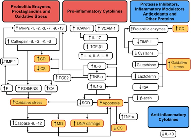
Keratoconus has been classically defined as a progressive, non-inflammatory condition, which produces a thinning and steepening of the cornea. Its pathophysiological mechanisms have been investigated for a long time. Both genetic and environmental factors have been associated with the disease. Recent studies have shown a significant role of proteolytic enzymes, cytokines, and free radicals; therefore, although keratoconus does not meet all the classic criteria for an inflammatory disease, the lack of inflammation has been questioned. The majority of studies in the tears of patients with keratoconus have found increased levels of interleukin-6 (IL-6), tumor necrosis factor-α(TNF-α), and matrix metalloproteinase (MMP)-9. Eye rubbing, a proven risk factor for keratoconus, has been also shown recently to increase the tear levels of MMP-13, IL-6, and TNF-α. In the tear fluid of patients with ocular rosacea, IL-1α and MMP-9 have been reported to be significantly elevated, and cases of inferior corneal thinning, resembling keratoconus, have been reported. We performed a literature review of published biochemical changes in keratoconus that would support that this could be, at least in part, an inflammatory condition.


Figures

Figure 1
Figure 1
The inadequate balance between pro-inflammatory cytokines, proteolitic enzymes, protease inhibitors, inflammatory modulators, and antioxidants may lead to an altered corneal structure and function in keratoconus, triggering a vicious circle between oxidative stress, keratocyte apoptosis, and increased activity of metalloproteinases., , On cell cultures of keratocytes from keratoconic corneas, basal PGE2 production was found to be 10 times greater than in normals. PGE2, whose release may be induced by TNF-α, has been shown to have inhibitory effects on collagen synthesis (CS) and to increase collagen degradation (CD). The tear film in keratoconus have shown increased levels of pro-inflammatory molecules: IL-1α, -4, -5, -6, -8, and -17, TNF-α, TGF-B1 (TGFβ-1), ICAM-1, and VCAM-1., , , , β-Actin gene has been found to be downregulated and the protein absent in corneal buttons from keratoconus patients. The elevated levels of IL-1-α and TNF-α, and low levels of β-actin have been related to triggering apoptosis of keratocytes., , , In keratoconus, levels of proteases such as lysosomal cathepsin-B, -G, -K, and -S, and metalloproteinases (MMPs) are elevated. IL-6 and TNF-α can stimulate the production of several MMPs (-1, -2, -3, -7, -9, and -13) and CATS., , , , , There is also a decrease in the levels of several antioxidant or anti-inflammatory molecules: SOD, glutathione, lactoferrin, IgA, and IL-10., , , , , , An important decrease in the level of protease inhibitors such as cystatins (inhibitors of cysteine proteases) and TIMP-1 (inhibitor of MMPs) have also been reported., , , , , The increased activity of several proteolytic enzymes results in higher concentrations of ROS, RNS, cytotoxic aldehydes (CAs) and peroxynitrates (Ps) (which decreases the activity of TIMP-1 and increase MMP-2),, , , , and given the lower production of SOD possibly related to IL-1α, an environment with high oxidative stress and low pH is formed, causing an increase in the activation of the caspases (caspase-9 and -12), mitochondrial dysfunction (MD), and DNA damage, which eventually lead to increased apoptosis. All of these could probably be the result of a complex interaction of both genetic predisposition and environmental triggering factors, such as eye rubbing and contact lenses wear (the ‘two-hit hypothesis') in keratoconus., , , , ,

Comment in

  • Reply.
    Shajari M, Kohnen T. Shajari M, et al. Cornea. 2017 Jan;36(1):e1-e2. doi: 10.1097/ICO.0000000000001053. Cornea. 2017. PMID: 27918358 No abstract available.

References

    1. Rabinowitz YS. Keratoconus. Surv Ophthalmol. 1998;42:297–319. - PubMed
    1. Krachmer JH, Feder RS, Belin MW. Keratoconus and related noninflammatory corneal thinning disorders. Surv Ophthalmol. 1984;28:293–322. - PubMed
    1. Davidson AE, Hayes S, Hardcastle AJ, Tuft SJ. The pathogenesis of keratoconus. Eye (Lond) 2014;28:189–195. - PMC - PubMed
    1. Brunsting LA, Reed WB, Bair HL. Occurrence of cataracts and keratoconus with atopic dermatitis. AMA Arch Derm. 1955;72:237–241. - PubMed
    1. Spencer WH, Fisher JJ. The association of keratoconus with atopic dermatitis. Am J Ophthalmol. 1959;47:332–344. - PubMed

MeSH terms