

A rare presentation of primary leiomyosarcoma of ovary in a young woman
- PMID: 25932048
- PMCID: PMC4406529
- DOI: 10.3332/ecancer.2015.524
A rare presentation of primary leiomyosarcoma of ovary in a young woman
Abstract
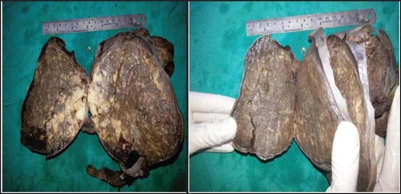
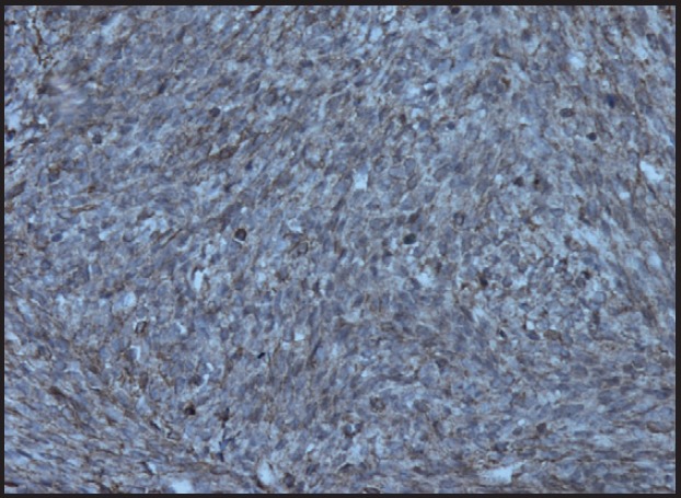
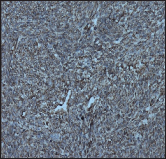
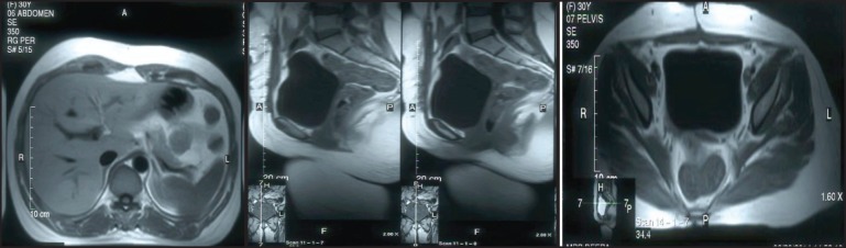
Primary ovarian leiomyosarcoma is a very rare tumour which is most commonly seen in postmenopausal women. A case is described here involving a 30-year-old female who presented with pain in the abdomen. The patient underwent laparotomy with peritoneal lavage, bilateral salpingo-oophorectomy, and total abdominal hysterectomy. Microscopic and immunohistochemical findings established the diagnosis of primary ovarian leiomyosarcoma. Similar to sarcoma of other tissues, an adjuvant chemotherapy regimen consisting of vincristine, epirubicin, and cyclophosphamide was given for this present case. Six months after treatment completion, the patient is on regular follow-up and disease-free on clinical and radiological examination.
Keywords: chemotherapy; leiomyosarcoma; ovary; radiation therapy; soft tissue sarcoma; surgery.
Figures

References
-
- Friedman HD, Mazur M. Primary ovarian leiomyosarcoma. An immuno-histochemical and ultrastructural study. Arch Pathol Lab Med. 1991;115(9):941–5. - PubMed
-
- Kurian RR, Preethi J, Remadevi AV. Leiomyosarcoma of ovary–a case report. Indian J Pathol Microbiol. 2005;48(1):19–20. - PubMed
-
- Rosai J. Female Reproductive System, Ovary. In: eds Ackerman’s Sur Pathol. 7. Washington DC: CV Mosby Co; 1989. p. 1163.
Publication types
LinkOut - more resources
Full Text Sources
Other Literature Sources
Medical
Molecular Biology Databases