

Expression of the P2Y2 receptor in the terminal rectum of fetal rats with anorectal malformation
- PMID: 25932095
- PMCID: PMC4402742
Expression of the P2Y2 receptor in the terminal rectum of fetal rats with anorectal malformation
Abstract
Objective: The expression and distribution of a subtype of purine receptors (P2Y2) in the terminal rectum of fetal rats with anorectal malformations (ARM) were examined to investigate their possible impact on the development of the enteric nervous system (ENS).
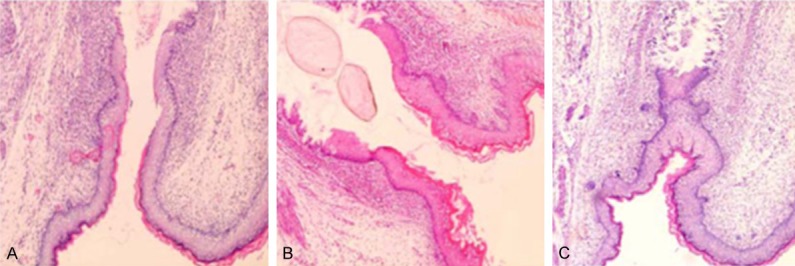
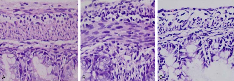
Methods: Pregnant Sprague-Dawley rats were randomly divided into a control group (5 rats) and an experimental group (20 rats). The experimental group was treated with ethylene thiourea (ETU). On gestational day 20, the intrauterine fetal rats were collected from both groups of pregnant rats. Sagittal sections of the pelvic perinea were stained with HE. P2Y2 protein and mRNA expression in the terminal recta of the fetal rats in the control group, the ARM group, and the ETU-treated group that exhibited no malformations (the ETU group) were detected by immunohistochemistry, western blot, and qRT-PCR.
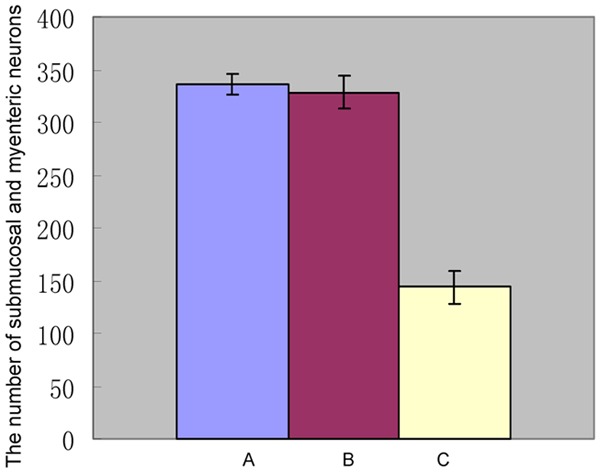
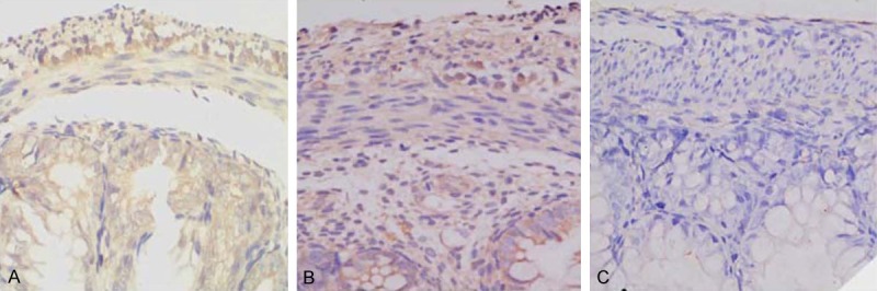
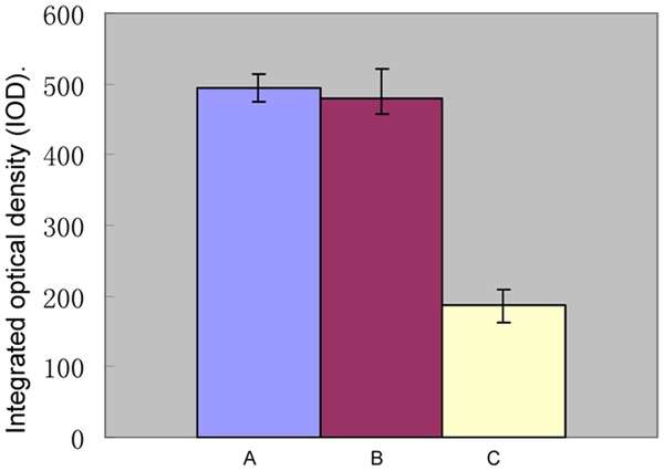
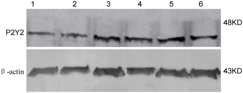
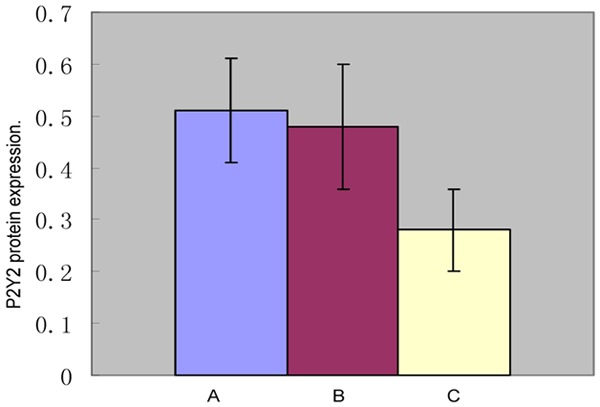
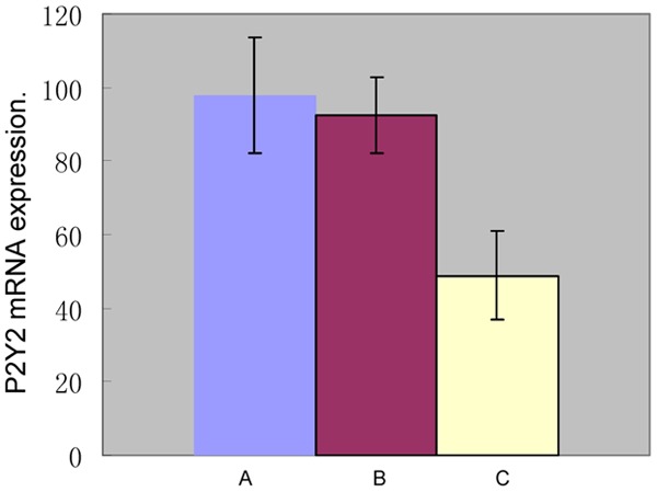
Results: The fetal rats in the control group showed normal position of the anal opening, with no malformation. The incidence of ARM was 89.2% for the fetal rats in the experimental group. The immunohistochemistry results showed that P2Y2 was expressed in the cytoplasm of the cells in the terminal rectum submucosa and myenteric plexus of the fetus rats in the control group, the ETU group, and the ARM group. The average integrated optical density (IOD) value for the ARM group was significantly lower than the IOD value for the control and ETU groups (186.48 ± 23.03 vs. 493.18 ± 19.70; 186.48 ± 23.03 vs. 479.48 ± 41.71, P<0.01), while the IOD value for the ETU group was comparable to the control group IOD (493.18 ± 19.70 vs. 479.48 ± 41.71, P = 0.360). The western blot and qRT-PCR results showed that the P2Y2 protein and mRNA expressions were significantly lower in the terminal rectum of the fetal rats in the ARM group than in the control and ETU groups (0.28 ± 0.08 vs. 0.51 ± 0.10, 0.28 ± 0.08 vs. 0.48 ± 0.12; 48.91 ± 12.17 vs. 98.03 ± 15.68, 48.91 ± 12.17 vs. 92.53 ± 10.43; P<0.01), while the P2Y2 protein and mRNA levels in the control group were comparable to the ETU group (0.51 ± 0.10 vs. 0.48 ± 0.12, P = 0.494; 98.03 ± 15.68 vs. 92.53 ± 10.43, P = 0.058).
Conclusion: P2Y2 may participate in and affect the development of ENS in the terminal rectum of fetal rats with ARM.
Keywords: Anorectal malformation; P2Y2; enteric nervous system; fetal rats.
Figures
References
-
- Gariepy CE, Mousa H. Clinical management of motility disorders in children. Semin Pediatr Surg. 2009;18:224–238. - PubMed
-
- Hamid CH, Holland AJ, Martin HC. Long-term outcome of anorectal malformations: the patient. Pediatr Surg Int. 2007;23:97–102. - PubMed
-
- Young HM, Newgreen D. Enteric neural crest-derived cells: Origin, identification, and differentiation. Anat Rec. 2001;262:1–15. - PubMed
LinkOut - more resources
Full Text Sources