

Three-D imaging of dental alveolar bone change after fixed orthodontic treatment in patients with periodontitis
- PMID: 25932177
- PMCID: PMC4402824
Three-D imaging of dental alveolar bone change after fixed orthodontic treatment in patients with periodontitis
Abstract
Objectives: The objective of this study was to radiographically quantify bone height and bone density in patients with periodontitis after fixed orthodontic treatment using cone beam computed tomography (CBCT).
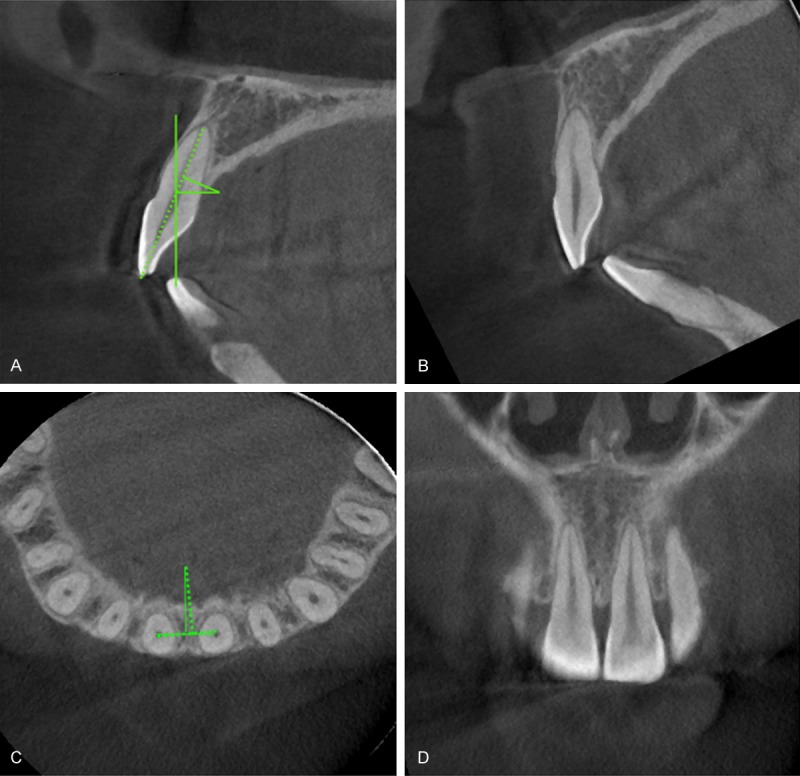
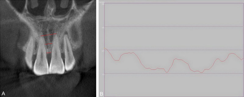
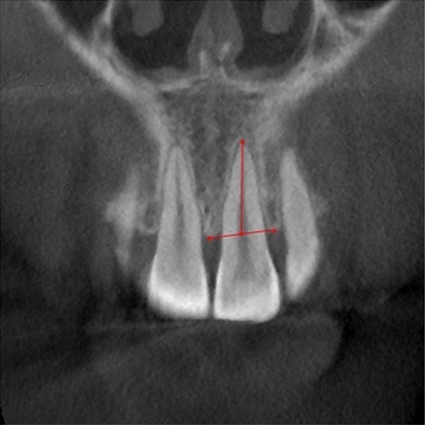
Materials and methods: A total of 81 patients including 40 patients with chronic periodontitis (group 1) and 41 patients with normal periodontal tissues (group 2) were selected. CBCT scanning for anterior teeth were taken before and after orthodontic treatment. Measurements of bone height and bone density were performed using CBCT software.
Results: The group 1 presented a statistically lesser bone density and bone height when compared to group 2 before treatment. There was a significant loss of bone density for both groups after orthodontic treatment, but bone density loss was significantly greater in the group 1. There was no statistically significant bone height change in two groups after treatment.
Conclusions: This study demonstrated that orthodontic treatment can preserve bone height but not capable of maintaining bone density, especially for patients with periodontitis. It is indicated that the change of bone density may be more susceptible than that of bone height when radiographically evaluating bone status under this combined periodontal and orthodontic therapy.
Keywords: CBCT; alveolar bone density; alveolar height; periodontal disease; tooth movement.
Figures
References
-
- Cao Z, Li C, Zhu G. MMP-1 promoter gene polymorphism and susceptibility to chronic periodontitis in a Chinese population. Tissue Antigens. 2006;68:38–43. - PubMed
-
- Wagenberg BD, Eskow RN, Langer B. Orthodontic procedures that improve the periodontal prognosis. J Am Dent Assoc. 1980;100:370–373. - PubMed
-
- Zachrisson BU. Clinical implications of recent orthodontic-periodontic research findings. Semin Orthod. 1996;2:4–12. - PubMed
-
- Tavares CA, Allgayer S, Calvete Eda S, Polido WD. Orthodontic treatment for a patient with advanced periodontal disease: 11-year follow-up. Am J Orthod Dentofacial Orthop. 2013;144:455–465. - PubMed
-
- Corrente G, Abundo R, Re S, Cardaropoli D, Cardaropoli G. Orthodontic movement into infrabony defects in patients with advanced periodontal disease: a clinical and radiological study. J Periodontol. 2003;74:1104–1109. - PubMed
LinkOut - more resources
Full Text Sources